Contact
New Bolton Center Kennett Square, PA
Emergencies & Appointments:
610-444-5800
Directions
Ryan Hospital Philadelphia, PA
Emergencies:
215-746-8911
Appointments:
215-746-8387
Directions

Leica SP5-II Confocal/Fluorescence
Lifetime Imaging Microscope


Description

The fully integrated PicoQuant fluorescence lifetime imaging system on this inverted laser scanning confocal microscope gives users the option of analyzing fluorescent probe distribution based on fluorescence lifetime in addition to fluorescence intensity.  This is particularly useful for examining dynamic protein-protein interactions by Förster resonance energy transfer (FRET), which can be measured as a shortening in the average fluorescence lifetime of the FRET donor.

Purchase of this instrument was made possible by NIH grant S10 RR027128-01.  Please acknowledge this funding source on any publications that include work performed on the Leica SP5 FLIM Confocal.

Applications

  •  Routine confocal microscopy and spectral scans
  • FLIM-based Förster resonance energy transfer (FRET)

Leica SP5-II FLIM

SP5 FLIM, Imaging Core

Technical Specifications: Leica SP5 FLIM tech sheet (PDF)

Scheduling Core Instruments

Registered users can access the PVIC online scheduler to reserve and use the Penn Vet Imaging Core instruments.

If you are not already registered, please contact the core manager.

Application Examples

Confocal Microscopy and 3D reconstruction of Z-stacks

This movie shows a 3D reconstruction of scanning laser confocal images of GFP-tagged Ebola VP40 protein (green) being expressed by an HEK293T cell. The cells were fixed then stained with HCS CellMask Deep Red (red).

Images were taken on the Penn Vet Imaging Core’s Leica SP5 II inverted confocal/FLIM microscope with a 100x oil immersion lens by scanning the sample with 488 nm Argon and 647 nm HeNe confocal lasers. Image Z-stacks were rendered into the 3D image shown here with Volocity software from Perkin Elmer, accessed through the core’s Volocity license server.

Image courtesy of Jonathan Madara, Gordon Ruthel, Bruce Freedman, and Ron Harty, Department of Pathobiology, UPenn School of Veterinary Medicine.


FLIM-based Förster Resonance Energy Transfer (FRET)
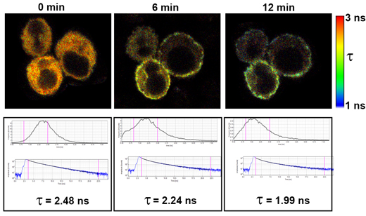
STIM+Orai FLIM-FRET, Imaging Core

The fluorescence lifetime of Cerulean fluorescent protein was measured at various times after treatment with thapsigargin in Jurkat cells transfected to express Cerulean-STIM1 and Venus-Orai1. Thapsigargin causes a depletion of ER Ca2+ stores, resulting in STIM1 relocalization to associate with Orai1 on the plasma membrane and form CRAC Ca2+ channels.

As Cerulean-STIM1 associates with Venus-Orai1, FRET occurs between Cerulean (the donor) and Venus (the acceptor), which results in a shortening in the average fluorescence lifetime of Cerulean. 

The images are fluorescence lifetime heatmaps based on the scale shown to the right. These images correspond to graphs of average lifetime, decay curves, and average Cerulean fluorescence lifetimes (τ). 

Fluorescence lifetime imaging was performed on the Penn Vet Imaging Core’s Leica SP5 II inverted confocal/FLIM microscope with a 63x water immersion lens by scanning the sample with a 405 nm pulsed diode laser.

Images courtesy of Xiaohong Liu, Gordon Ruthel, and Bruce Freedman, Department of Pathobiology, UPenn School of Veterinary Medicine.