Mary Avarbock
- Research Specialist & Laboratory Manager
- Email: avarbock@vet.upenn.edu
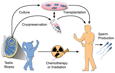
Male germline stem cell preservation. Before treatment for cancer by chemotherapy or irradiation, a boy could undergo a testicular biopsy to recover stem cells. The stem cells could be cryopreserved or, after development of the necessary techniques, could be cultured. After treatment, the stem cells would be transplanted to the patient's testes for the production of spermatozoa. The critical roadblock to widespread clinical use of this approach is the absence of culture techniques.
Clarence Freeman
Animal Research Technician

Rose Naroznowski
Animal Research Technician
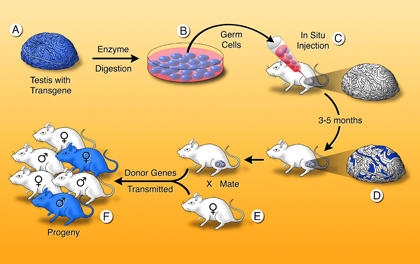
Testis spermatogonial stem cell transplantation method. A single-cell suspension is produced from a fertile donor testis (A). The cells can be cultured (B) or microinjected into the lumen of seminiferous tubules of an infertile recipient mouse (C). Only a spermatogonial stem cell can generate a colony of spermatogenesis in the recipient testis. When testis cells carry a reporter transgene that allows the cells to be stained blue, colonies of donor cell–derived spermatogenesis are identified easily in recipient testes as blue stretches of tubule (D). Mating the recipient male to a wild-type female (E) produces progeny (F), which carry donor genes. Genetic modification can be introduced while the stem cells are in culture.
Nilam Sinha, PhD
Research Associate & Postdoctoral Fellow
Email: nilams@vet.upenn.edu
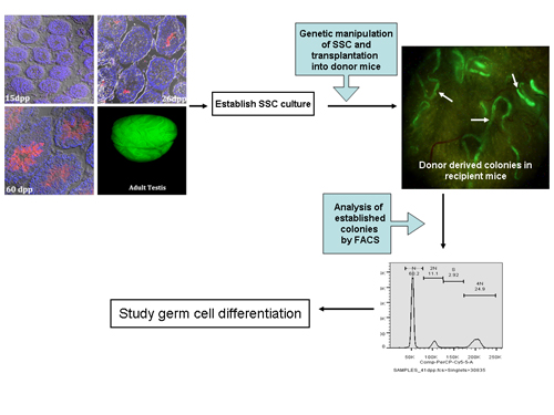
Studies on genes controlling spermatogenesis. Histological sections of a mouse testis showing germ cell differentiation and gradual appearance of spermatids and spermatozoa following birth (dpp is days postpartum) and an adult testis. In the histological sections (A,B,C), the red color represents the gradual appearance of protamine, which is present in spermatids and spermatozoa as they are produced following birth. The red stain is from an antibody for green florescent protein (GFP), which is expressed at the same time as protamine, because it is a transgene regulated by the protamine promoter and used as a marker for protamine in these experiments. Therefore, there is no red stain at 15 dpp (A), a small amount at 26 dpp (B), and in all spermatids and spermatozoa at 60 dpp (C), which exactly mimics the appearance of protamine in germ cells. In the adult testis (D), the green color is from GFP fluorescence under control of the protamine promoter. The variation in the intensity of the green in the adult testis represents the variation in the content of protamine at different stages of spermatogenesis in the adult testis. These mice, in which only spermatids and spermatozoa express GFP, are used in our experiments to study germ cell differentiation and determine the genetic regulation of spermatogenesis.
 Eoin Whelan, PhD
Postdoctoral Fellow
Email: ewhelan@upenn.edu
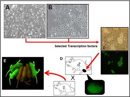
Reprogramming somatic cells to germ cells. Somatic cells, such as mouse embryonic fibroblast (A) and hair follicle stem cell (B) are being reprogrammed into spermatogonial stem cells through lenti-viral introduction of selected transcription factors. After successful reprogramming, converted cells should look morphologically similar to spermatogonial stem cells (SSC) in culture (C). The original cells taken from the mouse will have a fluorescence marker (GFP for example) linked to a promoter for a SSC specific gene. The cells will fluoresce upon successful conversion and when exposed to a specific wave length of light. Function of converted cells will be evaluated through transplantation (D). If colonization is extensive, the testis will have many fluorescent tubules, and the male can be mated to a female to produce fluorescent pups that contain the genes of the mouse from which somatic cells were grown (E). If the amount of spermatogenesis is small, for example a single colony, spermatozoa can be harvested and used to fertilize an oocyte by intra-cytoplasmic sperm injection. Gene modification or correction could be performed before conversion of the somatic cell to an SSC.
 Lauren Spring
Administrative Assistant & Office Manager to
R. L. Brinster, V.M.D., Ph.D.
Richard King Mellon Professor
of Reproductive Physiology
Department of Biomedical Sciences
School of Veterinary Medicine, 100E
University of Pennsylvania
3850 Baltimore Avenue
Philadelphia, PA 19104-6009
Telephone: 215-898-8805
Fax: 215-898-0667
Email: lspring@upenn.edu
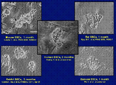
Appearance of spermatogonial stem cell cultures of rodent and non-rodent species. When grown in vitro, these stem cells from species separated by approximately 70 million years of evolution are remarkably similar in morphology. The mouse, rat and rabbit stem cells can be maintained in culture for longer than a year. However, stem cells of the baboon and human will grow for only about two months before disappearing. Nonetheless, the stem cells from these diverse species all require glial cell line-derived neurotrophic factor (GDNF) for their growth, indicating the extensive conservation of metabolic regulation in these stem cells. Experiments to understand the culture requirements of farm animals and primates are underway, including studies of genetic regulation of SSC self-renewal and spermatogenesis.
Oatley JM, Brinster RL. The germline stem cell niche unit in mammalian testes. Physiol Rev. 2012 Apr; 92(2): 577-595. PubMed Central PMCID: PMC3970841.
Wu X., Goodyear SM, Abramowitz LA, Bartolomei MS, Tobias JW, Avarbock MR, Brinster RL. Fertile offspring derived from mouse spermatogonial stem cells cryopreserved for more than 14 years. Human Reproduction. 2012 May; 27(5): 1249-1259. PubMed Central PMCID: PMC3329194.
Wu X., Goodyear SM, Tobias JW, Avarbock MR, Brinster RL. Spermatogonial stem cell self-renewal requires ETV5 mediated downstream activation of Brachyury in mice. Biol. Reprod. 2011 Dec; 85(6): 1114-1123. PubMed Central PMCID: PMC3223249.
Niu Z, Goodyear SM, Rao S, Wu X, Tobias JW, Avarbock MR, Brinster RL. MicroRNA-21 Regulates the Self-Renewal of Mouse Spermatogonial Stem Cells. Proc. Natl. Acad. Sci USA. 2011 Aug 2; 108(31): 12740-12745. PubMed Central PMCID: PMC3150879.
Kubota H, Wu X, Goodyear SM, Avarbock MR, Brinster RL. Glial cell line-derived neurotrophic factor and endothelial cells promote self-renewal of rabbit germ cells with spermatogonial stem cell properties. FASEB J. 2011 Aug; 25(8): 2604-2614. PubMed Central PMCID: PMC3136339.
Oatley JM, Kaucher AV, Avarbock MR, Brinster RL. Regulation of mouse spermatogonial stem cell differentiation by STAT3 signaling. Biol Reprod. 2010 Sep; 83(3): 427-33. PubMed Central PMCID: PMC2924805.
Wu X, Oatley JM, Oatley MJ, Kaucher AV, Avarbock MR, Brinster RL. The POU domain transcription factor POU3F1 is an important intrinsic regulator of GDNF-induced survival and self-renewal of mouse spermatogonial stem cells. Biol Reprod. 2010 Jun; 82(6): 1103-1111. PubMed Central PMCID: PMC2874496.
Wu X, Schmidt JA, Avarbock MR, Tobias JW, Carlson CA, Kolon TF, Ginsberg JP, Brinster RL. Prepubertal human spermatogonia and mouse gonocytes share conserved gene expression of germline stem cell regulatory molecules. Proc Natl Acad Sci USA. 2009 Dec 22; 106(51): 21672-7. PubMed Central PMCID: PMC2799876.
Oatley JM, Oatley MJ, Avarbock MR, Tobias JW, Brinster RL. Colony stimulating factor 1 is an extrinsic stimulator of mouse spermatogonial stem cell self-renewal. Development. 2009 Apr; 136(7): 1191-9. PubMed Central PMCID: PMC2685936.
Oatley JM and Brinster RL. Regulation of spermatogonial stem cell self-renewal in mammals. Annu. Rev. Cell Dev. Biol. 2008; 24: 263-286. PubMed Central PMCID: PMC4066667.
Oatley JM, Avarbock MR, Brinster RL. Glial cell line-derived neurotrophic factor regulation of genes essential for self-renewal of mouse spermatogonial stem cells is dependent on SRC family kinase signaling. J. Biol. Chem. 2007 Aug 31; 282: 25842-25851. PubMed Central PMCID: PMC4083858.
Brinster RL. Male germline stem cells: From mice to men. Science. 2007 Apr 20; 316: 404-405. DOI: 10.1126/science.1137741; PubMed PMID: 17446391.
Oatley JM, Avarbock MR, Telaranta AI, Fearon DT, Brinster RL. Identifying genes important for spermatogonial stem cell self-renewal and survival. Proc. Natl. Acad. Sci. USA. 2006 Jun 20; 103: 9524-9529. PubMed Central PMCID: PMC1480440.
Kubota H, Avarbock MR, Brinster RL. Growth factors essential for self-renewal and expansion of mouse spermatogonial stem cells. Proc. Natl. Acad. Sci USA. 2004 Nov 23; 101(47): 16489-94. PubMed Central PMCID: PMC534530.
Brinster RL. Germline stem cell transplantation and transgenesis. Science. 2002 Jun 21; 296: 2174-2176. DOI: 10.1126/science.1071607; PubMed PMID: 12077400.
Of the total publications, approximately 315 are in peer-reviewed journals and 84 are chapters/reviews. Included in the publications are 30 in Nature, 18 in Cell, 13 in Science, and 35 in Proc. Natl. Acad. Sci. USA. H-Index: 120
For a full list of publications, please visit PubMed
Ralph L. Brinster
Personal
Military Service:
1954-1956 Lieutenant, USAF
Office address:
School of Veterinary Medicine
University of Pennsylvania
Philadelphia, Pennsylvania 19104
Telephone: (215) 898-8805
Facsimile: (215) 898-0667
Email: brinster@vet.upenn.edu
Education
1949-53 B.S., School of Agriculture, Rutgers University
1956-60 V.M.D., School of Veterinary Medicine, University of Pennsylvania
1960-64 Ph.D. (Physiology), Graduate School of Arts & Sciences, University of Pennsylvania
Additional Training
1960 Postdoctoral Fellow, Jackson Laboratory, Bar Harbor, Maine, Summer.
1962 Postdoctoral Fellow, Marine Biological Laboratory, Woods Hole, Massachusetts, Summer.
Appointments
1960-64
Teaching Fellow, Department of Physiology, School of Medicine, University of Pennsylvania.
1964-65
Instructor, Department of Physiology, School of Medicine, University of Pennsylvania.
1965-66
Assistant Professor of Physiology, Department of Animal Biology, School of Veterinary Medicine, University of Pennsylvania, Philadelphia, USA.
1966-70
Associate Professor of Physiology, Department of Animal Biology, School of Veterinary Medicine, University of Pennsylvania.
1968-83
Program Director, Reproductive Physiology Training Program, School of Veterinary Medicine, University of Pennsylvania.
1969-84
Program Director, Veterinary Medical Scientist Training Program, School of Veterinary Medicine, University of Pennsylvania.
1970-
Professor of Physiology, School of Veterinary Medicine, University of Pennsylvania.
1970-
Professor of Physiology, Graduate School of Arts & Sciences and Graduate Faculty, University of Pennsylvania.
1975-
Richard King Mellon Professor of Reproductive Physiology, School of Veterinary Medicine and Graduate School, University of Pennsylvania.
1997-07
Scientific Director, Center for Animal Transgenesis and Germ Cell Research, School of Veterinary Medicine, University of Pennsylvania
2007-08
Co-Director, Institute for Regenerative Medicine, University of Pennsylvania.
Fellowships
1960-61
American Veterinary Medical Association Fellow, Graduate School Arts & Sciences, University of Pennsylvania.
1961-64
Pennsylvania Plan Scholar, Graduate School of Arts & Sciences, University of Pennsylvania.
Honors
New York Academy of Sciences Award in Biological and Medical Sciences, 1983.
Harvey Society Lecturer, 1984.
Member of the Institute of Medicine, National Academy of Sciences, 1986.
Fellow of the American Academy of Arts and Science, 1986.
Member of the National Academy of Sciences, 1987.
Honored (with L. Stevens) by an International Symposium at W. Alton Jones Cell Science Center (for pioneering work on development of the teratocarcinoma model and transgenic animals), 1987.
Fellow of the American Association for the Advancement of Science, 1989.
Distinguished Service Award of U. S. Department of Agriculture, 1989.
Invited Speaker at the Nobel Symposium on “Genetic Control of Embryonic Development”, 1991.
Pioneer Award from the International Embryo Transfer Society, 1992.
Juan March Foundation Lecture, Madrid, Spain, 1992.
Fellow of the American Academy of Microbiology, 1992.
Doctor Honoris Causa in Medicine, University of the Basque Country, Spain, 1994.
Charles-Léopold Mayer Prize (with R. Palmiter), The highest prize of the French Academy of Sciences (for development of transgenic animals), 1994.
Alumni Award of Merit, School of Veterinary Medicine, University of Pennsylvania, 1995.
First March of Dimes Prize in Developmental Biology (with B. Mintz) (for critical work in development of transgenic mice), 1996.
Carl Hartman Award, Society for the Study of Reproduction, 1997.
Bower Award and Prize for Achievement in Science, The Franklin Institute (for development of methods to transfer foreign genes into animals), 1997.
John Scott Award for Scientific Achievement, The City Trusts of Philadelphia, 1997.
Pioneer in Reproduction Research Award, The National Institute of Child Health & Human Development, National Institutes of Health, 1998.
Honored by a Special Festschrift Issue of the International Journal of Developmental Biology “Stem Cells and Transgenesis”, 1998.
George Hammel Cook Distinguished Alumni Award, Rutgers University, 1999.
Charlton Lecture, Tufts University School of Medicine, 2000.
Honorary Doctor of Science Degree, Rutgers, The State University of New Jersey, 2000.
Ernst W. Bertner Award, University of Texas M. D. Anderson Cancer Center (for research discoveries enabling development of transgenic animals), 2001.
Highly Cited Researcher (1980-2000), Designated by the Institute for Scientific Information, 2002. About 1 in 1000 authors are in this category.
Selected for the Hall of Honor, National Institute of Child Health and Human Development, 2003, (1 of 15).
Wolf Prize in Medicine, Israel (with M. Capecchi and O. Smithies) (for introducing and modifying genes in mice), 2003.
Gairdner Foundation International Award, Canada (for pioneering discoveries in germ line modification in mammals), 2006.
National Medal of Science, USA (for fundamental contributions to the development of transgenic mice), 2010.
International Society for Transgenic Technology Prize for outstanding contributions to the field of transgenic animals and stem cells, 2011.
Lifetime Achievement Award. From the Alumni of the University of Pennsylvania School of Veterinary Medicine, 2012.
Career Excellence in Theriogenology Award. From the Theriogenology Foundation on behalf of the American College of Theriogenologists and the Society for Theriogenology, 2012.
Honorary Doctor of Laws, University of Calgary, Canada, 2015.
Fellow of the American Physiological Society, 2015.