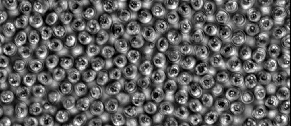
 The parasite Cryptosporidium, transmitted through water sources, is one of the most common causes of diarrheal disease in the world.
“We have been interested in the romantic life of the parasite Cryptosporidium for some time,” says Boris Striepen, a scientist in Penn’s School of Veterinary Medicine.
Cryptosporidium is a leading cause of diarrheal disease in young children around the world. The intestinal parasite contributes to childhood mortality and causes malnutrition and stunting. How a parasite like this one reproduces and completes its life cycle has significant impact on child health.
 Dr. Boris Striepen
“It’s the product of parasite sex, that is infectious agent here, a spore, that is transmitted by contaminated water,” Striepen says. “So, if you break its ability to have sex, you would break the cycle of transmission and infection.”
In a new paper in PLOS Biology, Striepen and colleagues in his lab tread new ground in understanding how Cryptosporidium reproduces inside a host. Using an advanced imaging technique allowed the scientists to observe the entire lifecycle in the laboratory. They found the parasite completes three cycles of asexual replication and then directly switches to male and female sexual forms. Their observations refute an intermediate stage that was introduced in the 1970s and align well with the original description of physician and parasitologist Edward Tyzzer who discovered this pathogen more than a century ago.
“What we have shown contradicts what you see in most textbooks today, including the description at the Centers for Disease Control and Prevention website,” Striepen says. “It’s really a super simple lifecycle that is completed in a single host in three days and only has three characters: asexual cells, male cells, and female cells.”
Other parasites, such as the malaria parasite Plasmodium, a “cousin” of Cryptosporidium, have more complicated and lengthy paths to follow an overall similar life cycle. While Crypto completes its lifecycle in one host, most malaria parasites move between two: a mosquito, where the parasite’s sexual reproduction occurs, and a human, where its asexual replication occurs.
“Cryptosporidium is a great model to study parasite development; you can see analogous steps to what happens with the malarial parasite, but it‘s much simpler because it all happens over only three days in one host, and we can observe it in simple cell cultures,” Striepen says.
In earlier work on Cryptosporidium, Striepen and colleagues had found that sexual reproduction appeared necessary for the parasite to move from one host to infect another but also to sustain itself in a host during chronic infection. Blocking developmental progression and parasite sex thus presents itself as a strategy to cure or prevent infection.
Cryptosporidium is a minuscule single-cell parasite that invades and reproduces within the cells of the intestine of its hosts. To get a closer look at what was happening, the researchers developed a live-cell microscopic imaging technique to track the progression of the parasite through multiple days in cell cultures. Using genetic engineering they added a fluorescent label to the nucleus of each parasite, allowing them to observe the replication of the parasite in real time and to distinguish its different lifecycle stages.
What they saw was the parasites “count to three,” says Striepen. Rather than responding to environmental cues, the parasites followed a rigid built-in plan. After infecting a culture, the parasite underwent three cycles of asexual reproduction. Each cycle took about 12 hours, during which the parasite established a home within the host cell and replicated itself resulting in eight new infectious parasites. Those were then released to infect surrounding host cells.
After these three waves of amplification, their fate changes abruptly, and they turn into either male or female gametes, or sex cells, in a process that also took about 12 hours. Tracking individual parasites and their offspring the researchers found no evidence for a specialized intermediate form assumed by many textbooks, demonstrating direct development.
Interestingly, the parasite appeared pre-committed to their future fate and carried that plan from one host cell into the next in a way not yet understood.
The researchers were intrigued to see that male and females arise from the infectious forms released from the same asexual parasites. “One of the really interesting aspects of sexual identity here is that it is not inherited and hard-wired in the genome but much more fluid,” Striepen says. “There’s an asexual cell that divides itself into genetically identical clones, and then those clones somehow become male or female on the fly, resulting in dramatically different cell shape and behavior.”
Future research will focus on the molecular mechanism of commitment to understand how this life cycle is programmed into the parasite’s biology. Understanding the life cycle of Cryptosporidium is critical in thinking about how to create a vaccine or therapy for the disease, Striepen says.
“How cells make decisions and execute developmental plans is one of the most fundamental questions in biology. Cryptosporidium offers a tractable system to better understand this mechanism in parasites. Hopefully we can gain insights that contribute to the understanding of cryptosporidiosis and malaria and lead the way to new urgently needed interventions for these important diseases.”
Boris Striepen is the Mark Whittier and Lila Griswold Allam Professor of Microbiology and Immunology at the University of Pennsylvania School of Veterinary Medicine.
Striepen’s coauthors were lab members Elizabeth D. English, Amandine Guérin, and Jayesh Tandel.
The study was supported by grants from the National Institutes of Health to Striepen and a postdoctoral fellowship from the European Molecular Biology Organisation to Guérin.