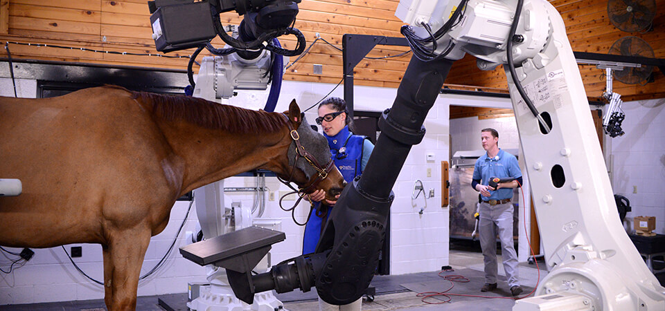
 Dr. Kyla Ortved, at left, and Dr. Kathryn Wulster
Arthritis can sideline a sport horse. Catching and treating the joint disease early is key to keeping an athletic equine comfortable and active. But when arthritis affects a horse’s elegant and powerful neck, with its complex map of muscles and vertebrae, it’s hard to pinpoint.
“Horses get arthritis in their necks much like people do,” said Dr. Kyla Ortved, Assistant Professor of Large Animal Surgery. “We’re recognizing it more frequently, but there’s not one defining symptom for the animal. Horses can have neurologic deficits, they can have neck pain, they can have lameness. These are the three things we see often. Sometimes arthritis is the cause, sometimes it’s not. But it’s often the default diagnosis because the disease is so hard to accurately diagnose.”
To improve arthritis diagnostics so horses can receive appropriate treatment before the disease advances too far, Ortved, who runs a lab focused on orthopedic disease, is teamed with Dr. Kara Brown, a resident in Equine Sports Medicine and Rehabilitation; Dr. Kathryn Wulster, Clinical Assistant Professor, Diagnostic Imaging; Dr. Elizabeth Davidson, Associate Professor of Sports Medicine and Sports Medicine Service Chief; and Dr. Amy Johnson, Assistant Professor of Large Animal Medicine and Neurology.
All experts in equine sports medicine, the group has just launched research into an innovative two-step strategy to better detect arthritis in the cervical articular process joint. Their approach employs state-of-the-art robotic imaging technology and synovial fluid testing.
“Cervical osteoarthritis is something we don't know a lot about right now,” said Brown. “As sports medicine clinicians, we are all interested in learning how to better help these horses, and I approached Dr. Ortved about getting a research project started. We routinely collect synovial fluid during ultrasound-guided injections when treating horses with neck arthritis, but haven’t until now used it for anything. Robotic CT is also something we’re using more and more, so we have two great resources at our fingertips, and we figured why not use them.”
Robotic Arms Take Better Pictures
Traditionally, x-rays (radiographs) are the most common tool for diagnosing arthritis. They’re good... but not great.
“X-rays give us a lot of information but can’t show everything,” Ortved explained. “The lower neck is a complicated piece of anatomy, with joints on either side of the vertebrae and a lot of muscle. Interpretation of this region is complicated and very often does not yield a definitive diagnosis.”
 New Bolton Center sports medicine technician Heather Hunchuck assists Finnegan, owned by Amy Lambert and provided by Pine Creek Sport Horses of Chester Springs, PA, during a robotic neck scan. Josh Benson, imaging technician, is in the background adjusting the arms.
The next best option is a traditional CT scan but the horse must be anesthetized. “Many people don’t want to put their horse through general anesthesia,” said Wulster. MRI is the the gold standard for evaluation of this region in other species, but imaging the base of the equine neck with MRI is not currently possible.
New Bolton Center offers a new CT option that provides the benefits of CT without the horse needing to be put under anesthesia. In 2016, Penn Vet became the first teaching hospital in the world to install EQUIMAGINE™, a robotics-controlled imaging system for standing horses.
“A traditional CT machine uses a fan beam in a donut shape that limits how much of the horse you can image,” Wulster said. “Our robotic system, which uses cone beam CT, can image all the way to the caudal cervical/cranial thoracic spine and to mid-radius.”
The system’s arms move around the horse, capturing images while the animal is awake, moving, and load-bearing. Because of its range of movement and high-resolution capabilities, robotic CT gives much more detail than x-rays do and can pick up changes in bone density earlier than a radiograph can.
Wulster pointed out that Penn Vet is in the early stages of understanding what the futuristic CT can—and cannot—do: “At this point, traditional CT is easier to interpret. Cone beam CT picks up extraneous or erroneous information that can be confused with a lesion. And the contrast resolution between tissues with different densities is better in a traditional CT image.”
But, she added, “Diagnostic imaging has advanced so much in 30 years. We’re lucky to have many different options for our patients. We have x-ray, conventional CT, standing CT, MRI, standing MRI, and ultrasound equipment. Our role as clinicians is to use the best modality for each individual case. For example, we know conventional CT is better than x-ray for diagnosing subchondral bone injury. And we are currently looking to prove that robotic CT is as effective as conventional CT and better than x-rays.”
Biomarkers as the Way Forward
Even with these advances in imaging technology and techniques, said Ortved, “it's hard to have a definitive diagnosis based on images alone.” In addition to using robotic CT, the research team is exploring backing up a preliminary image-based diagnosis by analyzing a horse’s synovial fluid for biomarkers of arthritis.
“When horses are diagnosed with arthritis they receive a steroid injection in the affected joint,” Ortved explained. “To inject the steroid, we use an ultrasound to watch the needle go into the joint. Then we aspirate fluid to confirm we’re in the joint. Now, instead of just throwing away that fluid, we're measuring different biomarkers in it.”
Existing research of humans and animals with arthritis has shown concentrations of inflammatory biomarkers in synovial fluid. The New Bolton Center team is testing the fluid they collect to see if these biomarkers are present in horses with evidence of arthritis and whether the levels of biomarker concentration correlate with the severity of arthritis.
What they learn can potentially transform how—and at what stage in the disease—horses are diagnosed. “If we can find a correlation between synovial fluid biomarkers and arthritis and diagnose horses using robotic CT, our field is going to do a much better job at identifying and helping horses afflicted with this disease,” said Ortved.