Our Research
X-Chromosome Inactivation
X-Chromosome Inactivation (XCI) is one of the best-characterized epigenetic phenomena where long noncoding RNAs are key players that regulate gene expression. Female mammals (XX) have two X-chromosomes, and one X is randomly chosen for transcriptional silencing in order to equalize the expression of X-linked genes compared to males (XY). Thus females are mosaic for X-chromosome expression, where a cell will express the maternal or paternal X. One great example of female X-mosaicism are calico cats.
The X-chromosome contains about 1000 genes with roles in development, reproduction, metabolism, and immunity. Mammalian females (including humans) are healthier and have longer life-spans than males. Females have better survival rates than males from illnesses caused by infectious diseases, injury, or sepsis. The X-chromosome plays an important role in the hyper-responsive female immune system because this chromosome has the highest density of immune-related genes. Females are also more susceptible to a number of autoimmune diseases, suggesting that the enhanced immune function may be related to the loss of self-tolerance.
The X-chromosome contains about 1,000 genes with roles in development, reproduction, metabolism, and immunity. Mammalian females (including humans) are healthier and have longer life-spans than males. Females have better survival rates than males from illnesses caused by infectious diseases, injury, or sepsis. The X-chromosome plays an important role in the hyper-responsive female immune system because this chromosome has the highest density of immune-related genes. Females are also more susceptible to a number of autoimmune diseases, suggesting that the enhanced immune function may be related to the loss of self-tolerance.
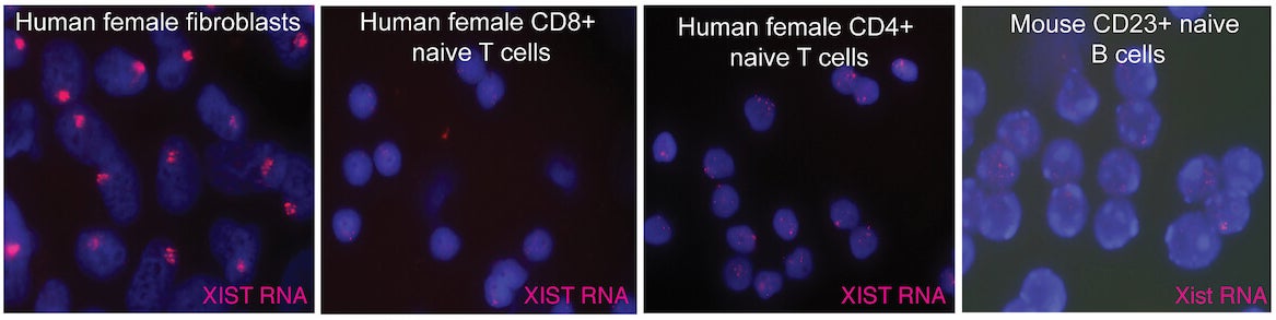
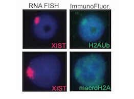
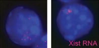
XCI occurs during early female embryonic development, and is initiated by expression of Xist RNA which recruits heterochromatic complexes for transcriptional silencing. Xist RNA and heterochromatin modifications associate with the inactive X chromosome with each cell division, and can be visualized using fluorescence microscopy. Xist RNA also functions to maintain XCI in somatic cells.
We recently discovered that XCI is maintained differently in female lymphocytes. Naïve T and B cells, which are quiescent, lack Xist RNA and heterochromatin marks on the inactive X. After lymphocyte stimulation, these epigenetic modifications re-localize back to the inactive X, as the cells re-enter the cell cycle and actively proliferate. This dynamic mechanism for XCI maintenance is unique to immune cells, and our lab investigates the molecular details of this process.
The major areas of investigation in our lab are:
- To understand how XCI is maintained in female lymphocytes, and how this contributes to female-biased autoimmunity;
- To investigate epigenetic mechanisms involving long noncoding RNAs during early human development and placental progenitors.

Calico cats, always female, visually demonstrate x-mosaicism (either mom’s or dad’s X is expressed) that arises from X-chromosome inactivation. The genesis for coat color are X-linked.
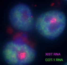
RNA FISH analysis of female hiPSCs using a probe for the long noncoding RNA XIST (pink), and COT-1 RNA (green) that detects nascent transcription. The XIST RNA., expressed from the inactive X, coats the entire chromosome. The inactive X is transcriptionally silent and resides in a COT-1 negative region.
Research Techniques
- Microscopy: Nuclear imaging of Xist RNA and the X-chromosome using RNA and DNA in situ hybridization (FISH); immunofluorescence detection of heterochromatic histone tail modifications
- Allele-specific transcriptional & epigenomic profiling of the inactive X and active X chromosome across somatic cell types (RNA-seq, CUT&RUN, ChIP-seq, Hi-C, DNA methylation)
- Mouse models of autoimmune disease, where animals spontaneously develop lupus-like disease symptoms. Induction of autoimmune disease features using pristane or imiquimod
- Human immune cell profiling of patients with female-biased autoimmune diseases, investigating epigenomic features of the X chromosomes and sex-biased differences with immune cell distribution and cell function
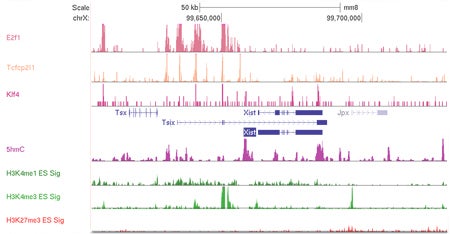
Epigenetic profile for the mouse Xic region, showing the locations for transcription factor binding and epigenetic modifications (peaks) for mouse embryonic stem cells. These modifications regulate gene expression at the Xic and are necessary for initiating X-chromosome inactivation during early development.
Reseach Projects
XCI maintenance is different in mammalian female lymphocytes
Human Naive T Cells
Human Activated T Cells
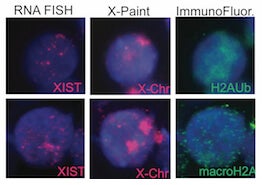
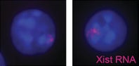
We have discovered that the inactive X chromosome lacks the typical heterochromatic modifications in female mature naïve T and B cells. This is the first example of physiologically-relevant female somatic cells that don’t have XIST/Xist RNA, H3K27me3, macroH2A, H2A-ubiquitin, and H4K20me modifications enriched on the inactive X. We also observed that in vitro activation of T and B cells stimulates the return of XIST/Xist RNA and some heterochromatic modifications back to the inactive X.
Using loss of function assays, we identified protein factors responsible for localizing XIST/Xist RNA back to the inactive X during lymphocyte activation. YY1 and hnRNP U, known XIST RNA binding proteins, return the XIST/Xist transcripts back to the inactive X. Elucidating the molecular mechanism of XIST/Xist RNA relocalization back to the inactive X, and how this transcript initiates the return of heterochromatin modifications, will reveal how the memory of transcriptional silencing is maintained after cell division in lymphocytes.
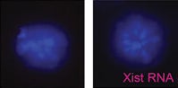
XIST/Xist RNA localization on the inactive X is perturbed in lymphocytes from female lupus patients and lupus mouse models
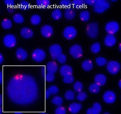
Healthy Female Activated T Cells
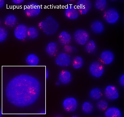
Lupus patient activated T cells
Remarkably, we have recently found that female lupus patients exhibit differences with XCI compared to healthy controls, which may explain the female bias of autoimmune disorders and why female lupus patients express higher levels of X-linked autoimmunity related genes compared to male patients. SLE-patient B cells have fewer typical XIST RNA clouds and more cells with dispersed patterns of XIST RNA or completely lacking XIST localization. Abnormal Xist RNA localization suggests partial reactivation of the inactive X-chromosome, which could increase gene expression. Using RNA FISH, we demonstrated that female lupus T and B cells frequently exhibit biallelic expression of CD40LG, CXCR3 and TLR7, which correlates with elevated expression of these genes. Moving forward, we are investigating causality of partial X-reactivation for lupus pathogenesis using mouse models. These will be the first experiments that directly connect transcription from the inactive X to lupus pathology.
Diversity of XCI maintenance mechanisms in immune cells
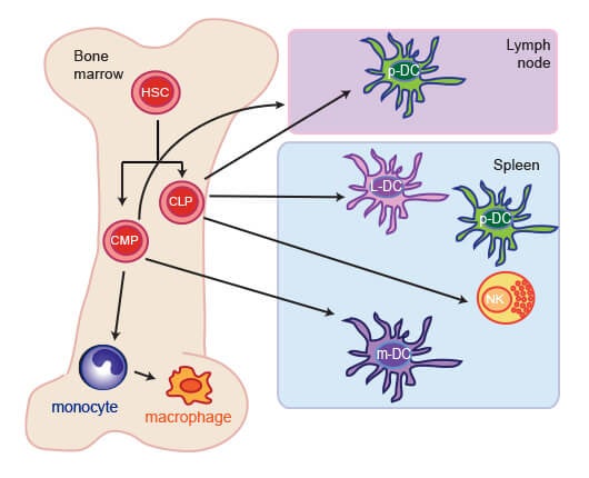
Our lab recently found that other immune cell populations, including dendritic cells, NK cells, and monocytes, have different mechanisms to maintain XCI. Using single-cell microscopy to image Xist RNA and heterochromatin modifications, we observed that monocytes and NK cells have faint Xist RNA ‘clouds’ and H3K27me3 foci, yet plasmacytoid dentritic cells (p-DCs) completely lack Xist RNA signals and enrichment of this histone mark. Unlike in humans, mouse p-DCs do not exhibit sex differences with interferon alpha production, and interferon signature gene expression in p-DCs is similar between males and females. Thus, immune cells use diverse (and unknown) mechanisms to maintain XCI, and we propose that these mechanisms are susceptible to error, thereby contributing to sex-linked autoimmune diseases. Our lab is actively investigating the molecular mechanisms that maintain XCI in a variety of immune cells.
NK-cells
Myleloid-DCs
Monocytes
Plasmacytoid DCs
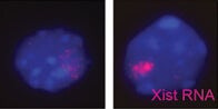
Lymphoid-DCs
Publications
“Impaired dynamic X-chromosome inactivation maintenance in T cells is a feature of spontaneous murine SLE that is exacerbated in female-biased models.” Jiwrajka N, Toothacre NE, Beethem ZT, Sting S, Forsyth KS, Dubin AH, Driscoll A, Stohl W, Anguera MC. (2023) J. Autoimmun. Sept; 139:103084. PMID: 37399593
“Unusual X chromosome inactivation maintenance in female alveolar type 2 cells is correlated with increased numbers of X-linked escape genes and sex-biased gene expression”. Sierra I, Pyfrom S, Weiner A, Zhao G, Driscoll A, Yu X, Gregory BD, Vaughan AE, Anguera MC. (2022) Stem Cell Reports. Dec 27:S2213-6711(22)00594-X. PMID: 36638790
“Long noncoding RNAs that function in nutrition: Lnc-ing nutritional cues to metabolic pathways.” Lovell, C. D. and Anguera, M.C. (2022) Annu Rev Nutr. 2022 Apr 18. PMID: 35436418
“The X in seX-biased immunity and autoimmune rheumatic disease”. Jiwrajka, N. and Anguera M.C. (2022) J Exp Med. Jun 6; 291(6):e20211487. PMID:35510951
“NHLBI Report: Impact of Sex and Gender on autoimmune lung disease, opportunities for future research”. Volkmann ER, Siegfried J, Lahm T, Ventetulo C, Mathai SC, Stenn V, Herzog E, Shansky R, Anguera MC, Danoff S, Giles JT, Lee YC, Drake W, Maier LA, Lachowicz-Scroggins M, Park H, Banerjee K, Fessel J, Reineck L, Vuga L, Crouser E, Feghali-Bostwick C. (2022) Am J Respir Crit Care Med. May 13. PMID: 35549658
“The dynamic epigenetic regulation of the inactive X chromosome in healthy human B cells is dysregulated in lupus patients”. Pyfrom S, Paneru B, Knoxx, J, Posso S, Buckner J, Cancro M, Anguera MC. (2021) Proc Natl Acad Sci. Jun 15;118 (24). PMID: 34103397
“Time to get ill: the intersection of viral infections, sex, and the X chromosome.” Forsyth, Katherine S. and Anguera, M.C. (2021) Curr Opin Physiol. Feb; 19:62-72. PMCID: PMC7553007
“Derivation and investigation of the first human cell-based model of Beckwith-Wiedemann syndrome”. Chang S, Hur SK, Naveh NSS, Thorvaldsen JL, French DL, Gagne AL, Jobaliya CD, Anguera MC, Bartolomei MS, Kalish JM. (2021) Epigenetics. Dec 16(12):1295-1305. PMID: 33300436