Bellwether Magazine
Discover Bellwether, Penn Vet’s premier biannual publication. Explore exciting stories and insights, and don’t miss the chance to delve into our extensive Bellwether Archives!
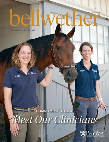
Featured Article
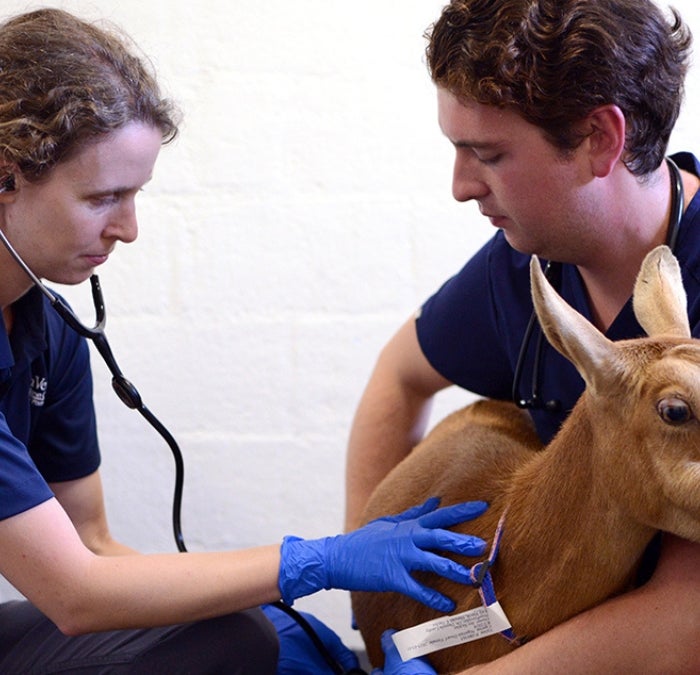
The Draw of Internal Medicine
Joy Tomlinson, VMD and Daniela Luethy’s Love of the Practice
Featured Articles
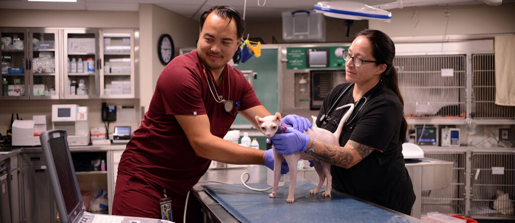
Ryan Hospital Staff Veterinarians Talk Life in Emergency Services and Critical Care
Drs. Catalina Montealegre and Charles Garneau-So participate in a Q&A.
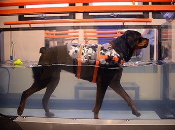
Ryan Hospital Launches Canine Hydrotherapy Services
Addition of Underwater Treadmill Expands Treatment Options for Arthritis, Rehabilitation
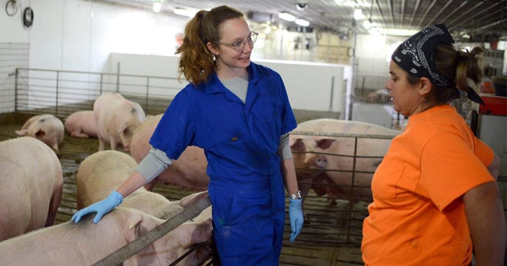
Tara Gaab, V’17
From Classroom to Farms to Back Again
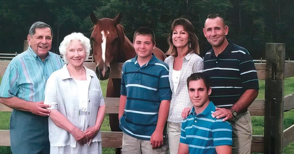
A Family Affair
Robert Marookian supports New Bolton Center in honor of his mother and brother
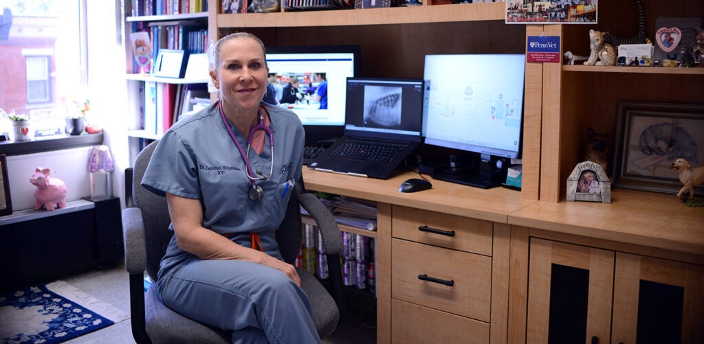
In the Office with Dr. Deborah Silverstein
Get to know Professor of Emergency & Critical Care Deborah Silverstein.