van Eps Laminitis and Endocrinology Laboratory
The van Eps Laminitis and Endocrinology Laboratory at New Bolton Center is focused on understanding the key events that drive laminitis under different circumstances in order to develop reliable means of prevention and treatment.
About Us
The van Eps Laminitis and Endocrinology Laboratory at New Bolton Center is dedicated to improving the prevention and treatment of laminitis through basic and applied (clinical) research. Our laboratory also offers a clinical endocrinology service, with rapid turnaround blood testing for insulin, ACTH and other endocrine biomarkers that can help identify laminitis risk and guide treatment.
We take a multidisciplinary approach to the study of laminitis, which includes:
- Developing and refining tests and novel biomarkers of endocrine dysfunction, the most common cause of laminitis in horses and ponies.
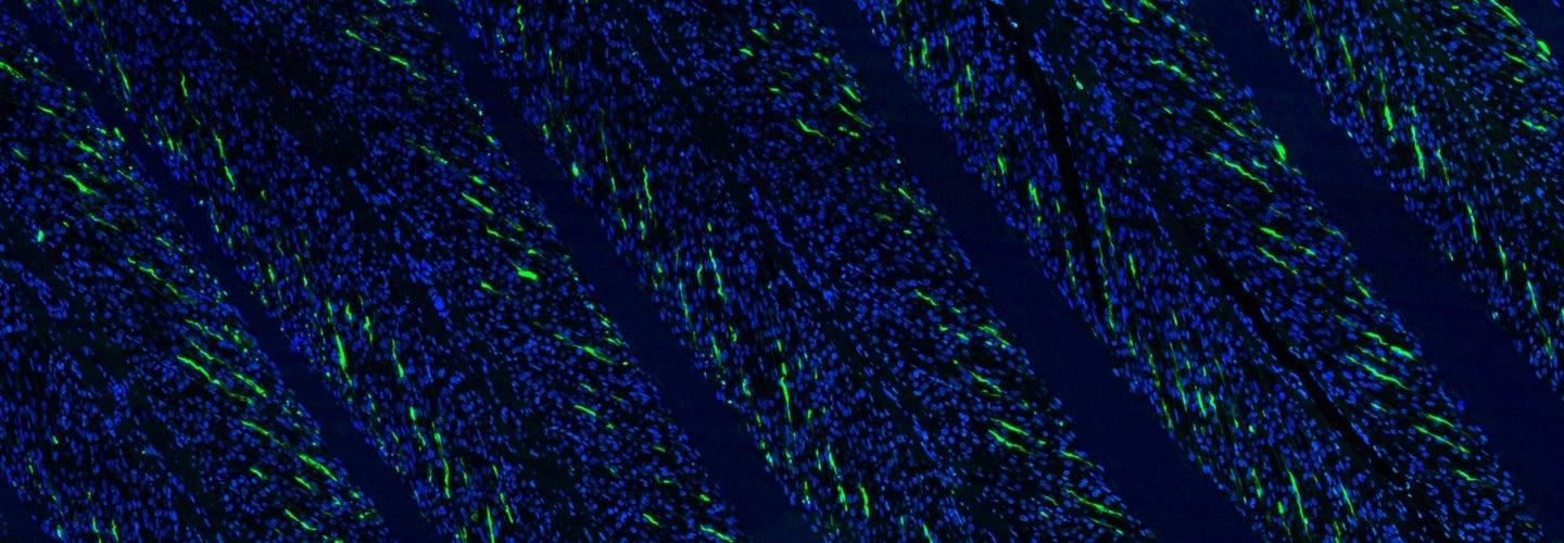
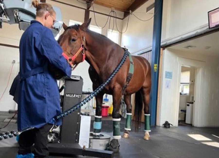
- Advanced imaging and sensor-based techniques to evaluate structure and function of the foot in health and disease
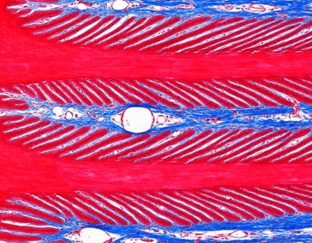
- Molecular techniques to examine events at a tissue level
- Biomechanical testing to study mechanical function both in vivo and ex vivo
Our Research
Laminitis occurs as a consequence of different primary problems in the horse and can be divided into three main categories based on cause:
- Endocrinopathic (hyperinsulinemia-associated) laminitis
- Sepsis-related laminitis
- Supporting-limb laminitis
Assay Services
We offer a range of diagnostic equine endocrine tests for clinical use.
Available Diagnostic Tests
Individual Assays
- Insulin
- Insulin monitoring (5 samples)
- ACTH
- Progesterone
- Adiponectin
- Bile acids
- Triglycerides
Combination / Panels
- Insulin and ACTH
- Insulin and adiponectin
- Adiponectin and leptin
- Endocrine panel (add leptin)
- Insulin dysregulation panel (add leptin)
- Insulin dysregulation panel with GGT and bile acids

Director, van Eps Laboratory
Andrew van Eps, BVSC, PhD, DACVIM
Professor, Equine Musculoskeletal Research
Join Us Today
At the van Eps Laminitis and Endocrinology Laboratory, we are always seeking highly motivated students and post-doctoral fellows with an interest in:
- Biomechanics and orthopedics
- Endocrinology (pancreatic and pituitary)
- Epithelial cell biology and the effects of inflammatory, ischemic, mechanical and growth factor signaling events on epithelial tissue homeostasis
Find Us
University of Pennsylvania
School of Veterinary Medicine
New Bolton Center
382 West Street Road
Kennett Square, PA 19348