Dentistry & Oral Surgery
What we do
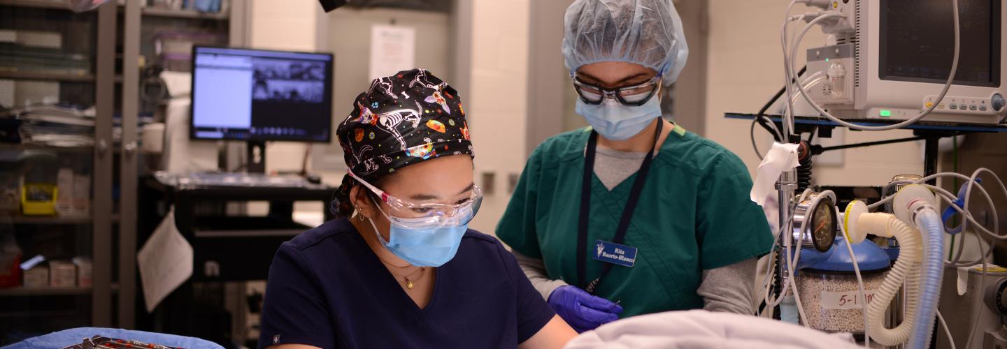
The Dentistry & Oral Surgery Service diagnoses and treats a wide variety of oral diseases in dogs, cats, and various special species. Most procedures are performed under general anesthesia, and all patients receive a thorough physical examination, including necessary diagnostic tests. This ensures that your pet receives the best care for dental and oral surgical procedures.
Our Services
Conditions and Procedures
Companion animals are affected by a wide range of oral conditions during their life stages, many of which are treatable. At Penn Vet’s Dentistry and Oral Surgery Service, our supportive team treats all conditions, including periodontal disease; stomatitis; endodontic disease in fractured, luxated, and avulsed teeth; oral tumors; malocclusions; and cleft palate defects.
Periodontal Disease
Periodontal disease is one of the most common diseases found in dogs and cats, affecting 60 to 70 percent of these animal populations, beginning as early as three years of age. This disease is an inflammatory response to infection of the periodontal tissues caused by dental plaque.
Periodontal disease occurs in two stages: (a) gingivitis and (b) periodontitis. Since gingivitis affects only the ginigiva — or gums — it is the reversible form of periodontal disease. When left untreated, however, the dental plaque toxins increase, causing a more severe inflammation of gingival tissues. At that point, the gingiva detach from the tooth, creating a periodontal pocket. Bacterial toxins and enzymes from inflammatory cells further destroy the periodontal tissues that surround the tooth, changing the disease from gingivitis to periodontitis. This progression can result in alveolar bone loss (alveolar bone is the bone that surrounds the tooth). The loss of alveolar bone is typically irreversible, and with increasing bone loss, the tooth becomes mobile and is eventually lost.
One of the most common signs of periodontal disease noted by pet owners is halitosis, or bad breath. Depending on the severity and progression of periodontitis, your animal may experience changes in eating habits, face rubbing, drooling, and weight loss.
Diagnosing and treating periodontal disease typically requires an oral examination that is conducted under anesthesia. The exam can be followed by professional dental cleaning, along with capturing dental radiographs of the patient’s mouth.
Fractured and Worn Teeth
Found in both dogs and cats, fractured and worn teeth are not uncommon injuries. Teeth are worn down either by malocclusion, in which the tooth-to-tooth contact is misaligned, or — in the case of dogs — by excessive chewing of tennis balls. Tooth fractures result when excessive force causes a tooth to break.
Once there is a fracture in a tooth, bacteria can enter directly into the pulp cavity or through a worn crown. Left untreated, the pulp becomes inflamed and necrotic, and a periapical abscess (abscess at the base of the tooth) may develop. Treatment options vary, depending on whether the pulp is exposed. If it is, either an extraction or endodontic therapy would be recommended. Dental radiographs provide the opportunity for a complete diagnosis and confirmation of treatment.
While this is not a true dental emergency that needs immediate care, the tooth should be evaluated within a reasonable time.
Luxated and Avulsed Teeth
Tooth displacement injuries frequently occur after automobile accidents, falls from great heights, fighting with other animals, or when an animal gets a tooth caught in something like a fence.
Luxation
A tooth is considered to be luxated when it is dislodged from its normal location. A tooth can be luxated to the side, into the alveolus (nasal cavity) in the case of a maxillary canine tooth, or to the outside.
Avulsion
Alvusion means that a tooth is completely out of its alveolar socket (space in which the tooth sits). In dogs, the teeth most commonly avulsed are the incisor and canine teeth.
Luxated and avulsed teeth are dental emergencies because these require immediate repositioning, stabilization, and endodontic therapy due to the likely loss of blood supply to the pulp. Successful replantation is influenced by the speed with which the tooth is replanted.
An avulsed tooth should be placed in a transport medium (such as fresh milk which keeps periodontal ligament cells vital for up to 6 hours) until arrival of the patient and tooth at our hospital.
Antibiotic therapy (amoxicillin/clavulanic acid, clindamycin, or doxycycline) must be started as soon as possible. The tooth is then replanted, and a splint is applied.
In one to two weeks, the splint is removed, and endodontic therapy is performed with a calcium hydroxide paste fill. In another two weeks, standard root canal therapy is performed. Resorption of the replanted tooth is a common complication, particularly if the avulsed tooth has been allowed to dry in air for longer than 20 minutes.
Oral Tumors
Oral tumors may be benign or malignant (having the potential to spread to distant sites). Even those that are benign may be locally invasive and may result in oral pain, bleeding, drooling, or difficulty eating.
Unfortunately, dogs and cats often show no obvious clinical signs until the oral tumor is already very large. It is important to have your animal receive a thorough oral examination on a regular basis so that, if tumors occur, these can be diagnosed in the early stages. To determine tumor type and treatment options, an incisional biopsy of the tumor may be conducted prior to treatment. Treatment may include one or more of the following:
- Surgery: Removal of a portion of the lower (mandibulectomy) or upper jaw (maxillectomy) is generally very well tolerated, and in many cases, result in pets with good function and cosmetic appearance.
- Radiotherapy: Radiotherapy (radiation therapy) can be useful, either alone or in combination with surgery, depending on the tumor type, location, and the ability to remove the tumor surgically.
- Chemotherapy: Few oral tumors respond to chemotherapy alone, but chemotherapy may be prescribed to minimize spread to distant sites.
- Immunotherapy: Recent advances in vaccine technology offer a completely new treatment approach for certain tumor types.
Patients with rapidly growing oral tumors should receive immediate veterinary medical attention. Oral tumors are best treated before they have a chance to enlarge to the point where they are no longer surgically removable.
Dental Procedures
Our services offers a wide range of oral care for your pet.
- Oral Examination
- Dental Radiographs
- Computed Tomography Imaging
- Dental and Periodontal Cleaning
- Periodontal Surgery
- Tooth Extraction
- Endodontic Therapy
- Restorative Dentistry and Prosthodontics
- Orthodontics
- Oral and Maxillofacial Trauma Management
- Temporomandibular Joint Surgery
- Oral and Maxillofacial Cancer Surgery
- Facial Reconstruction
- Palate Surgery
- Salivary Gland Surgery
- Oral Medicine
For Our Clients
If your animal experiences a dental emergency, we can help. Knowing what comprises an emergency is helpful, and gaining a better understanding of what we can do and what support you can provide your pet can make all the difference.
Traditionally, our Dentistry and Oral Surgery Service clinicians have not only taken care of patients with dental problems, but are also responsible for the care of patients with oral and maxillofacial disease due to infection, cancer, or trauma. Certain emergencies affecting the teeth, mouth, and face require immediate veterinary medical attention. These include but are not limited to:
- Very recent tooth fractures if there is interest in saving the tooth, the animal should be put on antibiotics until referral to the veterinary dentist or oral surgeon.
- Sharp or blunt head trauma injuries, including lip and tongue lacerations, and oral bleeding.
- Tooth luxations and avulsions are true dental emergencies. Put the animal on antibiotics and place avulsed tooth in milk until referral to the veterinary dentist or oral surgeon.
- Mandibular and maxillary swellings associated with oral and maxillofacial tumors.
- Swellings around the nose, mouth, jaws, face, and neck associated with inflammation / infection.
- Jaw fractures, temporomandibular joint luxations, symphysis separations, acute palate defects.
- Acute inability to open or close the mouth.
Emergency cases admitted through our Emergency Service receive primary attention and will undergo immediate treatment as feasible. Contact Emergency Service at (215) 746-8911.
An effective home oral hygiene routine for pets includes daily tooth brushing, appropriate diets, and various oral health care products like chlorhexidine-based rinses, chew toys, and dental treats.
Tooth Brushing
Brush your pet’s teeth daily using a soft-bristled, appropriately-sized toothbrush and pet-safe toothpaste. Do not use toothpaste for humans, as it can upset your pet’s stomach and pose health risks if swallowed.
Dental Health Diets
Certain veterinary diets may be advantageous for maintenance of oral health. Many manufacturers have considered the relationship of diets to oral health of pets. So-called ‘dental diets’ were designed to either mechanically or chemically reduce plaque and/or calculus (tartar) accumulation. Some products provide a combination of both actions. The mechanical action is derived from a larger than usual, hard kibble that fractures into few large pieces as it is penetrated by the tooth, rather than crumbling into many tiny pieces. The large pieces are penetrated again, and thus the more crunching that is performed, the more abrasive action results and the bacteria-laden plaque is being disturbed. Some kibble also has layers of different textures that contribute to plaque disruption. Diets that help to clean teeth by chemical action are coated with various chemicals that have been shown to reduce the accumulation of plaque or calculus. Polyphosphates are used in some of these diets.
Toys and Dental Treats
Chew toys and dental treats can be an important part of any pet dental health program. Toys and treats should be used in combination with daily tooth brushing, oral health care products, appropriate diets, yearly dental check-ups, and professional dental cleaning and periodontal therapy as necessary.
There are many different chew toys and dental treats commercially available for our pets. They should not be too hard, as very hard materials can fracture teeth.
- Acceptable toys include soft stuffed animals, flexible rubber bones, soft plastic balls, and ropes. Please note that the toy should be appropriate for the size of the animal, and caution should be exerted when pets are left unobserved when playing with toys.
- Inappropriate toys and treats include plastic bones made of hard nylon, meat bones (cooked and uncooked), and cow hooves. Rocks and large ice cubes can also fracture teeth and should be avoided. Tennis balls are a popular toy for many dogs; however, they are very abrasive to teeth because they collect tiny particles of dirt and sand and will wear down the teeth and occasionally cause pulp exposure.
Specific breeds, such as Yorkshire terriers and miniature schnauzers are more prone to developing periodontal disease; therefore, a professional dental cleaning should be performed more frequently.
The frequency of the need for professional dental cleanings is dependent upon several factors. If thorough home oral hygiene is being provided on a daily basis, the bacterial accumulations should be minimal, and scaling and polishing procedures can be performed less frequently.
Your pet should have an annual oral examination performed by a professional to document the presence of abnormal conditions such as periodontal disease, fractured or decayed teeth, tumors, ulcers, etc.
Professional dental cleanings require your pet to be anesthetized in order for the skilled and trained operator to remove debris from below the margins of the gums (subgingivally). Since periodontal disease causes the destruction of the supporting structures of the teeth (gingiva, periodontal ligament, and bone), cleaning the crowns of an awake dog without addressing what lies beneath only provides a cosmetic benefit. This superficial procedure does not address the disease in deeper tissues or less accessible sites.
In general, the condition (color, texture, shape) of the gingival tissues will dictate the need for placing your pet under general anesthesia to have professional dental scaling, polishing and intra-oral radiographs.
In order to perform a thorough periodontal examination, dental radiography, scaling and polishing, gingival curettage and root planing, the pet must be under general anesthesia. Anesthetic gas and oxygen are delivered through an endotracheal tube, thus ensuring pain-free procedures and also protecting the airways from aspirating fluids or debris. Owners of pets naturally are concerned when anesthesia is required for their pet. However, anesthesia-free dentistry performed by untrained individuals is inappropriate for several reasons, including:
- Significant safety concerns for the patient and operator.
- Insufficient cleaning of inaccessible tooth surfaces.
- No debridement of periodontal pockets.
- Oral discomfort and serious pain.
- Accidental aspiration of debris that can result in pneumonia and death.
Furthermore, it is illegal for anybody but licensed veterinarians or supervised and trained veterinary technicians to practice veterinary medicine.
Although anesthesia will never be 100% risk-free, modern anesthetic and patient evaluation techniques used in veterinary hospitals minimize the risks, and millions of dentistry and oral surgery procedures are safely performed each year.
Our Care Team
Veterinarians


Residents

Shannon Heintz, VMD

Robert Thompson, VMD
Staff
Jeanette Eliason, CVT, RDH, VTS
Veterinary Nurse Supervisor, Dentistry
Brianna O’Brien, CVT
Research
The research program is focused on clinical research in dentistry and oral surgery. Our primary goals are to prevent disease and to optimize patient care. This can be accomplished by: enhancing knowledge of oral pathology development and, expanding diagnosis and treatment options.
Clinical research makes use of existing patient material by maintaining comprehensive medical and dental records, collecting specimens (tissue and fluid samples), and ensuring adequate follow-up. Investigational studies on privately-owned animals are sometimes performed (e.g., on patients with oral cancer when available treatment options have failed or are declined by the owner). These require consent by the patient owner, and must be approved by the University of Pennsylvania Institutional Animal Care and Use Committee (IACUC).
Continuing Education
The following continuing-education opportunities in dentistry and oral surgery are provided by members of the Dentistry and Oral Surgery Service other qualified instructors:
- Observatory rotation
- Laboratory courses
- Preparation for the AVDC or EVDC entry examination
Observatory Rotation
This program was established for visiting veterinary technicians and veterinarians who are interested in continuing clinical education in dentistry and oral surgery. Visitors will be observers of clinical and surgical cases but cannot have primary case responsibility. This service is provided on mutually convenient dates and on a space-available basis. The applicant is to provide an agenda of items and define specific goals that are to be met during their stay at the Ryan Veterinary Hospital (e.g., visitation time, extent of Diplomate involvement, certain procedures to be observed, credentials requirements to be met, etc.). The costs vary depending on the specific requirements.
Laboratory Courses
The Dentistry and Oral Surgery Service offers various laboratory courses to veterinary professionals. The four to eight-hour classes consist of one or more didactic presentations and hands-on practice. The classes are limited to four to six participants and are offered several times per year.
Ryan Veterinary Hospital
Emergencies:
(215) 746-8911
By Appointment:
(215) 746-8387