Interventional Radiology
What we do
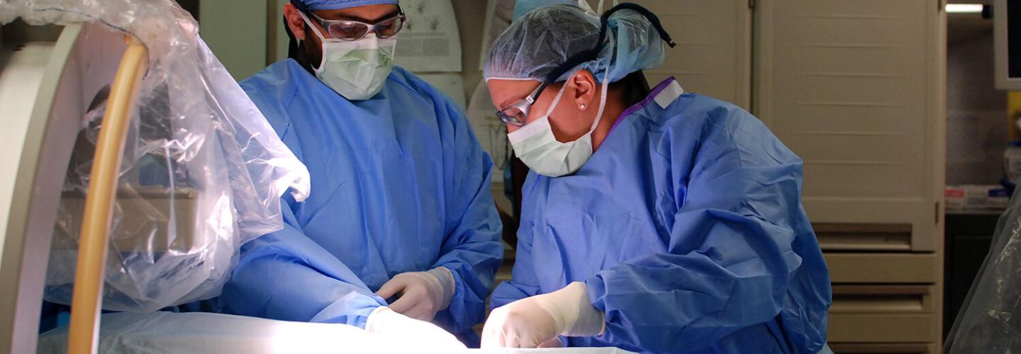
Interventional radiology (IR) involves the use of fluoroscopy and specialized equipment to gain access to different structures in the body, and deliver materials for diagnostic and therapeutic purposes.
IR techniques are minimally invasive, and can lead to fewer negative outcomes, shorter anesthesia times, and brief stays. Interventional radiology can be applied to patients of all sizes. We are leading the way in training the next generation of specialists this new and growing field.
Our Service
Interventions
Our Service
Interventions
Respiratory Interventions
- Nasopharyngeal Stensosis
- Tracheal Stenting
- Tracheal Foreign Body Retrieval
Gastrointestinal Interventions
- Esophageal Balloon Dilation and Esophageal Stenting
- Esophageal-jejunal Feeding Tubes
- Colonic Stenting
Biliary Interventions
- Endoscopic Retrograde Cholangiopancreatotgraphy (ERCP) and Biliary Stent Placement
- Laparoscopic Cholecystostomy Tubes
Urincary Techniques – Kidney & Ureter
- Ureteral Stenting
- Percutaneous Nephrolithotomy (PCNL)
- Percutaneous Nephrostomy Tube Placement
- Cystoscopic-Guided Ectopic Ureter Laser Ablation
- Ureteroscopy for Idiopathic Renal Hematuria
- Extracorporeal shock-wave lithotripsy (ESWL) for Nephro/Ureterolithiasis
- Urinary Bladder and Urethra laser lithotripsy
- Urethral Stenting for Malignant Obstructions
- Antegrade Urethral Catheterization
- Percutaneous Cystostomy Tubes
Our Care Team
Veterinarians


Intern and Staff

Lara Derr, CVT, LTS (Emergency Care)
Interventional Radiology Veterinary Nurse
Ryan Veterinary Hospital
Emergencies:
(215) 746-8911
By Appointment:
(215) 746-8387