Penn Vet Researchers Evaluate a New Device That Could Streamline Vision-Saving Therapies
Retinal blindness caused by the death of photoreceptors, the light-sensing cells within the retina, affects millions of people worldwide. Novel gene and cell therapies to prevent or rescue vision loss are currently being developed and tested. These treatments often need to be delivered directly to the eye and specifically under the retina, which is a delicate surgical procedure that requires a vitreoretinal surgeon’s expertise and dexterity. Now, vision scientists at the University of Pennsylvania School of Veterinary Medicine (Penn Vet) have shown that a novel, clinically used surgical device could make that task safer, more controlled, and potentially more effective in a preclinical model.
In a recently published study in Drug Delivery and Translational Science, a team of vision scientists from the Division of Experimental Retinal Therapies (ExpeRTs) at Penn Vet, led by William A. Beltran, DVM, PhD, DECVO, have worked with a group of scientists and drug delivery engineers to demonstrate that the Orbit® Subretinal Delivery System (Orbit® SDS), a novel surgical tool used previously in gene and cell therapy clinical trials (NCT02659098, NCT02286089, NCT03846193), can place therapies directly beneath the retina using a minimally invasive approach in a large-animal model. By accessing the treatment site from the back of the eye rather than through its center, the device avoids the need to puncture the retina itself—a step that can lead to complications such as inflammation, bleeding, or unintended leakage of therapeutic material.
“This represents a significant evolution in how we think about delivering treatments to the subretinal space,” says William Beltran, the Corinne R. and Henry Bower Professor of Ophthalmology and Director of the Division of ExpeRTs, and senior author of the study. “We showed that it’s possible to reach the retina with greater control and potentially less trauma.”
Using an established large-animal model with eye anatomy closely resembling that of humans, the researchers tested both clinical-grade (Orbit® SDS 2nd Generation) and prototype versions of the device customized for preclinical animal models in a range of eye sizes—including those comparable to human neonates. In nearly all cases, they were able to guide a flexible cannula through a natural tissue plane in the back of the eye and inject gene therapy material beneath the retina with high precision.
“Precision is everything when it comes to treating the retina,” says Jennifer Kwok, a veterinarian and postdoctoral fellow in the Beltran Lab and lead author of the study. “Even small improvements in how therapies are delivered can make a big difference in safety and success.”
One of the major goals of the study was to evaluate a persistent issue in subretinal surgery: preventing the backflow—or reflux—of gene therapy vectors into unintended parts of the eye. While the team was able to avoid leakage into the vitreal cavity of the eye — a central gel-filled space located in front of the retina and a common site of reflux with standard techniques—they did observe some escape of vector material along the cannula’s path in the suprachoroidal space. Although confined and not associated with serious side effects at lower doses, this finding underscores the importance of optimizing delivery parameters to reduce off-target exposure and inflammation.
“Our results highlight both the promise of this route for gene therapy delivery and the care needed to fine-tune it,” says Beltran. “Even when the surgical pathway is improved, how and where the therapeutic material ends up traveling to still matters, as release to the wrong locations of the eye can trigger inflammation, particularly when using devices optimized for clinical use in a preclinical animal model.”
The study also explored how eye anatomy affects device performance. For example, in eyes with a reflective structure called the tapetum — a feature not found in humans but common in many animal species — advancing the device’s needle could be more difficult. To overcome this, the team developed custom needle designs that improved delivery in such regions, offering a blueprint for adapting the device for use in veterinary patients.
The findings add to growing momentum in the field of ocular gene and cell therapies, where improved delivery methods could make treatments safer, more scalable, and more effective. As researchers continue to refine these approaches, tools like the Orbit® SDS could play a critical role in expanding access to vision-restoring therapies.
The work was conducted in collaboration with Raghavi Sudharsan, PhD, a research assistant professor in experimental ophthalmology at Penn Vet, investigators at the University of Florida (Shannon Boye, Stanford Boye), and at Genentech, Inc. (Kirsten Stoner Cummiskey, Tom Meyer, and Sam Reichenbacker).
The work was supported by the Foundation Fighting Blindness Center and TRAP grants, the National Eye Institute (grants RO1-EY006855, RO1-EY17549, and P30-EY001583).
Related News

Wild birds are driving the current U.S. bird flu outbreak (link is external)
Since late 2021, a panzootic, or “a pandemic in animals,” of highly pathogenic bird flu variant H5N1 has devastated wild birds, agriculture, and mammals.
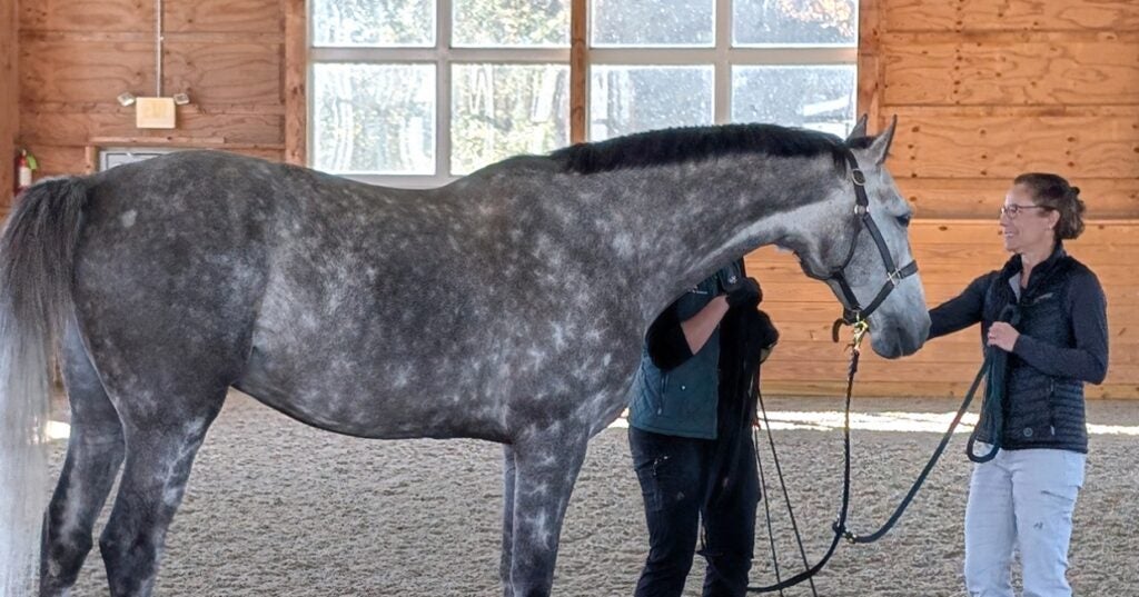
Behind the Breakthroughs: Amy Johnson
Balancing clinical care with scientific inquiry, Penn Vet’s Amy Johnson leads efforts to decode the complexities of neurologic diseases in horses
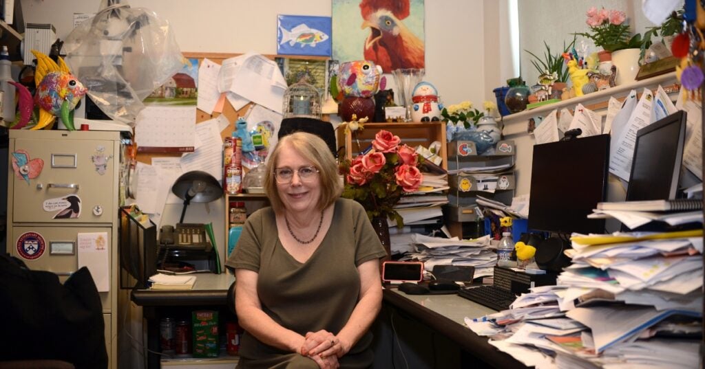
In the Office with Donna Kelly, DVM, MASCP, DACPV, DACVPM
Donna Kelly, DVM, MASCP, DACPV, DACVPM, shares her New Bolton Center office with the campus’s microbiology reference library.
About Penn Vet
Ranked among the top ten veterinary schools worldwide, the University of Pennsylvania School of Veterinary Medicine (Penn Vet) is a global leader in veterinary education, research, and clinical care. Founded in 1884, Penn Vet is the first veterinary school developed in association with a medical school. The school is a proud member of the One Health initiative, linking human, animal, and environmental health.
Penn Vet serves a diverse population of animals at its two campuses, which include extensive diagnostic and research laboratories. Ryan Hospital in Philadelphia provides care for dogs, cats, and other domestic/companion animals, handling more than 30,000 patient visits a year. New Bolton Center, Penn Vet’s large-animal hospital on nearly 700 acres in rural Kennett Square, PA, cares for horses and livestock/farm animals. The hospital handles more than 6,300 patient visits a year, while our Field Services have gone out on more than 5,500 farm service calls, treating some 22,400 patients at local farms. In addition, New Bolton Center’s campus includes a swine center, working dairy, and poultry unit that provide valuable research for the agriculture industry.