Diagnostic Imaging
What we do
We offer a comprehensive selection of diagnostic imaging modalities and a cross-disciplinary team of board-certified faculty, residents, interns, and specialized technical staff to manage the care of your animal.
Our Services
What We Offer
Below is a list of imaging options that our service offers.
- Digital radiography
- Fluoroscopy with a high-speed radiographic camera
- Computed tomography (CT)
- Magnetic resonance imaging (MRI)
- Nuclear medicine
- Nuclear scintigraphy
- Positron emission tomography (PET)
- NaF and FDG radiotracers
- New Bolton Center’s board-certified radiologists review the scans as they are performed and are available to assist in image acquisition, when needed.
- All images are reviewed by the clinician on the case, as well as a board-certified radiologist, with reports available within 48 hours.
For information or to schedule an appointment, please call (610) 925-6198 or email sportmed@vet.upenn.edu.
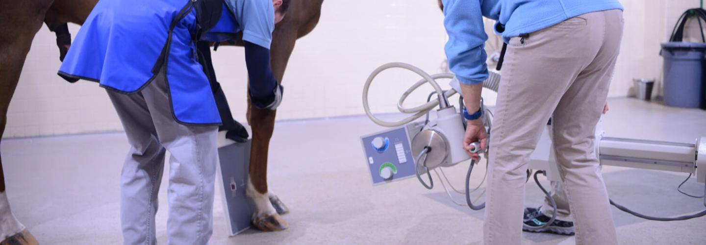
Digital Radiography
An 800 mA X-ray generator in the radiology suite provides imaging for standing, adult large animals; covering head, neck, chest, shoulders, elbows, and stifles. This generator can be used to image regions of the horse that can be difficult to image in the field, including the caudal cervical spine, thoracolumbar spine, pelvis, and abdomen. Myelograms can also be performed on the cervical spine under general anesthesia.
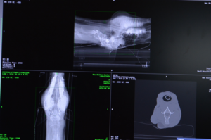
Computed Tomography (CT)
Computed tomography (CT) can be used to evaluate bony and soft tissue lesions in 3-D, helping to identify abnormalities that are incompletely seen or even missed on standard radiographs. CT is most useful for evaluating bone.
What We Offer
- Robotic imaging of the head, cervical spine and distal limb (mid-radius to foot and distal tibia to foot)
- Esophagrams / TVEC fluoroscopy
The Use of CT Scans During Surgery
At New Bolton Center, the integration of intra-operative computed tomography (CT) has transformed the way we approach orthopedic surgery. This advanced imaging technology allows our surgical team to visualize fractures and other abnormalities in precise detail and in three dimensions, before, during, and after surgery. By accurately identifying the full extent of an injury, CT enhances surgical planning and improves outcomes.
Many of these procedures are now performed using minimally invasive techniques, which rely on small incisions rather than fully opening the limb. Intraoperative CT, along with real-time fluoroscopy (continuous X-ray imaging), plays a critical role in guiding these delicate interventions with accuracy and confidence.
Robotic CT Imaging
Penn Vet was the world’s first veterinary teaching hospital to use a robotics-controlled imaging system for computed tomography, which can be used to image the horse without general anesthesia.
This system has proven value for both clinical and research applications.
Obtaining CT scans with New Bolton Center’s robotic imaging system offers unique advantages:
- The system enables our radiologists to diagnose conditions difficult to detect with other imaging modalities.
- The patient is awake and standing, unencumbered by an enclosed gantry.
- Scan time is reduced compared to CT scans that require general anesthesia.
- Obtaining the scans with sedation instead of anesthesia saves time and money, and decreases risk to the patient.
- The modality produces high-quality, multi-planar reconstructions and 3D images.
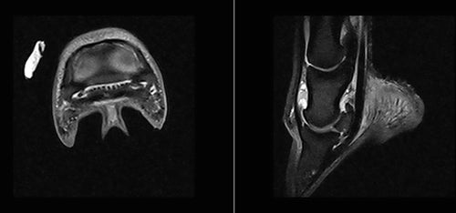
Magnetic Resonance Imaging (MRI)
Magnetic Resonance Imaging (MRI) uses a magnetic field and radio waves to produce highly detailed images of the body, offering exceptional evaluation of both soft tissue and bone. Compared to radiography, computed tomography (CT), and ultrasound, MRI provides superior soft tissue contrast—making it especially valuable for evaluating complex lameness cases.
O-Scan MRI
The O-Scan is a low-field MRI system designed specifically for imaging the distal limbs of the horse—from the hoof to the carpus (knee) or tarsus (hock). Scans are performed under general anesthesia to ensure image quality and patient safety.
MRI excels in detecting soft tissue injuries, such as tendon and ligament damage, but also identifies subtle bone pathology that may not be visible with other imaging techniques. This makes it an essential tool in the early diagnosis and management of equine orthopedic conditions.
Available Studies
Foot, Bilateral feet
- The single-foot imaging includes a limited study of the opposite foot or a limited study of the pastern
Proximal suspensory, Bilateral suspensory
- Proximal suspensory includes a limited study of the opposite suspensory
Fetlock, Bilateral fetlock
Carpus, Bilateral carpus
Tarsus, Bilateral tarsus
Appointments are available Monday through Friday, to schedule an appointment, please call (610) 925-6198 or email sportmed@vet.upenn.edu.
Nuclear Medicine
Nuclear medicine, commonly referred to as nuclear or bone scintigraphy, involves the administration of a radiopharmaceutical that binds to bone where there is injury, or at sites of active bone formation. Used in conjunction with lameness examination and diagnostic analgesia, this modality can assist in identifying the cause of lameness.
Potential Candidates for Nuclear Scintigraphy
Horses that might benefit from nuclear scintigraphy include:
- Horses with suspected stress related bone injury
- Horses in which radiographic images are negative or confusing
- Horses in which pain causing lameness can be localized using nerve blocks but the cause of pain cannot be determined on radiography and ultrasonography
- Horses with high-speed lameness, or poor performance
- Horses with numerous, subtle lameness abnormalities
- Horses with neck, back, or pelvic problems
- Horses with suspected soft tissue injury such as suspensory ligament, skeletal muscle, or soft tissue structures in the navicular region
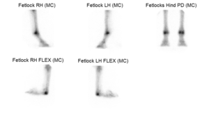
Bone scans should be performed in conjunction with a thorough lameness examination, which can be performed by your veterinarian or here at New Bolton Center. The results are then interpreted along with all other clinical and diagnostic information. Horses must remain in the hospital for 24 hours after radiopharmaceutical administration to allow the radioactivity to subside.
Positron Emission Tomography (PET)
Positron Emission Tomography (PET) is a form of nuclear medicine imaging, similar to scintigraphy, that allows veterinarians to detect subtle changes in bone and soft tissue metabolism. While PET scans originally required general anesthesia, advances in technology now allow the procedure to be performed safely with standing sedation.
Both PET and scintigraphy are used to identify areas of increased activity—or “hot spots”—that may signal early or microscopic injury, often before structural damage becomes visible on other imaging modalities. This is especially important in the detection and prevention of limb fractures, which can be life-threatening in horses. Early diagnosis through PET plays a critical role in protecting equine athletes and improving outcomes.
What to Expect
A small dose of a radioactive agent is injected approximately 30 minutes prior to imaging. This agent distributes through the body and accumulates in regions with increased bone turnover. Once the horse is sedated, the procedure takes between 3-5 minutes to image each site. In less than 30 minutes, both front feet and both front fetlocks can be imaged.
Results
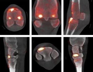
(Top row) CT/PET fused images of a fetlock in a horse demonstrate many sites of uptake of the radioactive agent in the fetlock indicating multiple sites of bone injury
(Bottom row) CT/PET fused images of a carpus (knee) in a horse demonstrate significant uptake of the radioactive agent in the third carpal bone indicating bone injury
Clinical Applications
Specialists at New Bolton Center use imaging to enhance their diagnostic work-up. Applications include:
Soft Tissue Surgery and Dentistry
- Evaluation of the head, including the teeth and paranasal sinuses.
- Learn more about dentistry.
Orthopedic Surgery
- Evaluation of trauma and wounds
- Lameness work-up
- Surgical planning
- Find out more about surgery.
Sports Medicine
- Lameness and poor performance work-up
- Learn more about the sports medicine service.
Neurology
- Cervical spine imaging
- Learn more about neurology.
Cardiovascular Medicine
- Evaluating the heart and lungs in patients with cardiac disease
- Find out more about cardiology.
Internal Medicine
- Evaluating the lungs in horses with respiratory illness
- Screening the abdomen for colonic sand or intestinal stone formation in horses with colic
- Read about internal medicine.
Translational Opportunities
Penn Vet researchers and clinicians collaborate with colleagues at Penn Medicine, Nemours Children’s Health System, and other human medical centers about possible applications of new imaging systems.
Translational applications include:
- Motion correction technology used to image standing, sedated horses is similar to what is used to image infants and pediatric patients
- Diagnosis and serial monitoring of naturally-occurring musculoskeletal disease including injuries that affect tendons, ligaments, and bone
- Intra-operative imaging techniques that can be performed during surgery
Ultrasonography
Ultrasound is an excellent method to evaluate soft tissue structures and the surface of bone. Ultrasound is known as a “user dependent modality” and our faculty have advanced training in musculoskeletal ultrasound and general ultrasound imaging. We have several state of the art “console” or hospital units in addition to portable capabilities. In our clinic, ultrasound is routinely but not exclusively used for evaluation for soft tissue injury in the lame horse, causes of acute or chronic colic, weight loss, thoracic disease, neonatal foals, high risk pregnant mares and our farm animal species. It is used to perform ultrasound guided injections, biopsies and intra-operative procedures, thus improving the accuracy and safety of these procedures over ‘blind’ techniques.
For information or to schedule an appointment, please call (610) 444-5800. Appointments may be made by the owner, trainer, or referring veterinarian.
Our Care Team
Service Chief, Radiology
Kathryn W. Bills, VMD, DACVR, DACVR-EDI
Associate Professor of Clinical Large Animal Diagnostic Imaging
Clinicians
Our clinicians come from various backgrounds, including expertise in cardiac, CT, MRI, radiology, ultrasonography, and robotic imaging, ensuring comprehensive and advanced diagnostic services.


Virginia B. Reef, DVM DACVIM, DACVSMR


Residents

Andrew Collins, MVB, MRCVS

Julia Fox, DVM

Natalie Goolik, DVM, DACVR

Maryam Iranmanesh, DVM

Romina Landazuri, DVM

Robb Kessel, VMD
Rocio Remolina-Palomino, DVM, MsC

Yu (May) Wang, DVM

Charmaine Tam, BVMS

Robert Wise, DVM, DACVR

Jin Yu, DVM
Staff
Carole Johnson, RTR
Director, Imaging & Clinical Service
Sports Medicine & Imaging
Katie Minacci
Supervisor of Sports Medicine and Imaging Technicians
Josh Benson
Advanced Imaging Technical Specialist
Jess Harris
Imaging Technician
Juliette Hopkins
Imaging Technician
Dannielle Snyder
Imaging Technician


