Core Laboratories
A history of
Connecting Research
Our research facilities include state-of-the-art core laboratories on both Philadelphia and Kennett Square campuses, and offers researchers throughout the University of Pennsylvania the opportunity to use highly specialized technology, and benefit from the expertise of world-class pathologists.
Animal Model Core & Comparative Orthopedic Research Laboratory
The Lab is part of the Institute for Medical Translation at Penn Vet New Bolton Center. It functions as an ecosystem for medical translation and multidisciplinary collaboration providing a bridge…
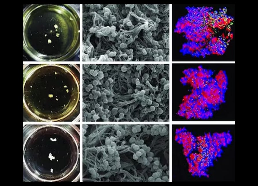
The Center for Host-Microbial Interactions
The Center for Host-Microbial Interactions (CHMI) is a shared resource for high-throughput sequencing and data analysis.
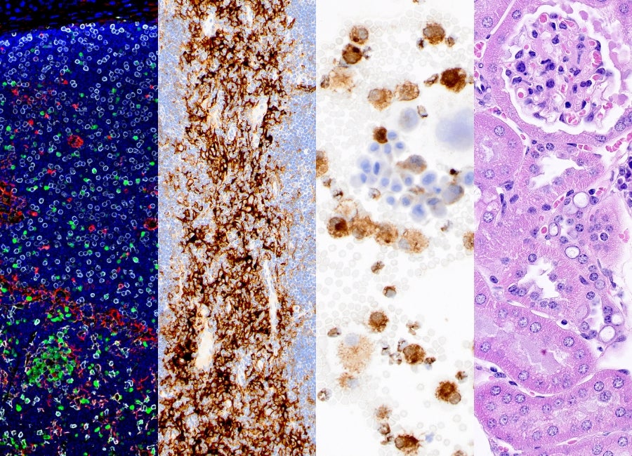
Comparative Pathology Core
The core provides expert pathological characterization and validation of mouse and other animal models used in biomedical research.
Extracellular Vesicle Core
The (EV) Core Facility provides comprehensive or selected services in the necessary isolation, quantification and characterization of EVs.
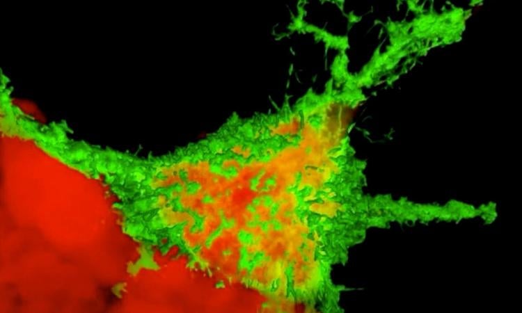
Imaging Core
The Penn Vet Imaging Core (PVIC) provides access to cutting-edge optical imaging capabilities for researchers.
Transgenic Mouse Core
A state-of-the-art fee-for-service facility that offers a full line of embryological manipulation services, focused on, but not limited to, murine model systems.

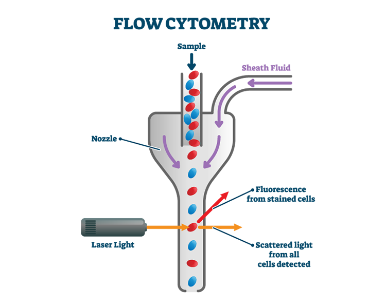
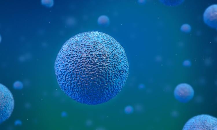
Flow Cytometry Core
Penn Cytomics is the sole flow cytometry shared resource laboratory (SRL) at the University of Pennsylvania. Our facility has 35+ instruments, which include analyzers and cell sorters, several of which have spectral and imaging capabilities. We also have a dual-fluorescence cell counter/viability instrument and a tissue dissociator for cell preparation.