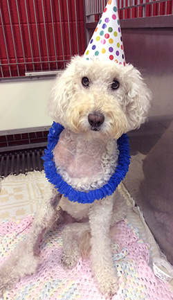
Chloe presented to Penn Vet’s Comprehensive Cancer Care service in June 2015 for an evaluation of several new masses. The nine-year-old female spayed Labradoodle with a history of mast cell tumors was evaluated by our expert team of medical, surgical, and radiation oncologists. Together, they determined that surgery should be the first approach for Chloe.
 Chloe was scheduled to have her masses excised in mid-June. Along with multiple low-grade/grade II mast cell tumors (MCTs), she had two high-grade/grade II MCTs located on her right medial elbow (completely excised) and right palmar carpus (incompletely excised).
High-grade tumors are aggressive tumors that can be invasive and recur locally. They also tend to spread to regional lymph nodes and may spread more distantly to organs such as the liver and spleen. In order to determine the extent of the spread of cancer, staging tests are performed, including aspirates of the draining lymph nodes and abdominal ultrasound.
Because high-grade tumors behave aggressively, adjuvant treatment is recommended following surgery. There are two arms to adjuvant treatment. The first is additional local-regional treatment consisting of radiation to the incision sites to prevent recurrence as well as prophylactically to the draining lymph nodes. Even if the lymph nodes are negative at staging, most dogs will fail in the nodes, so it is important to treat this site. The second is systemic treatment chemotherapy with vinblastine and prednisone.
It was determined that Chloe would receive both radiation therapy and concurrent chemotherapy. In early July 2015, a CT scan was obtained to aid in planning for Chloe’s radiation therapy, and she was started on her first dose of chemotherapy. It is important that the CT be performed at the facility where the radiation will occur so that a member of the radiation oncology team can be present to ensure proper positioning of the patient.
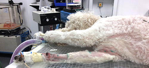
By mid-July, Chloe was set to start her 16 daily radiation treatments. Radiation therapy is given over a course of several weeks to help protect the normal tissues adjacent to the tumor by spreading out the total dose of radiation. Because patients are required to be anesthetized for treatment, we utilize a subcutaneous venous access port (SVAP), which is placed via a minor surgical procedure using minimally invasive techniques. The use of a SVAP ensures a simple, non-stressful method to obtain venous access during anesthesia and minimizes the need to use multiple veins, preserving these vessels for future use (such as blood draws and chemotherapy administration).
 Chloe completed her course of radiation therapy with only mild side effects and continued on with her chemotherapy. Her SVAP was left in place for blood sampling to minimize the stress of additional venipunctures. She completed her chemotherapeutic protocol by mid-September and her SVAP was removed.
At Penn Vet, we take great pride in this team approach. The medical, surgical, and radiation oncology teams round together twice a day and work side-by-side to provide the most appropriate, comprehensive, and timely care for patients. Read more about Penn Vet’s Comprehensive Cancer Care service.
We encourage telephone consultations to help facilitate your patient’s initial oncology appointment:
For radiation oncology, please call 215-573-3317.
For medical and surgical oncology, call 215-746-8387 and ask to transfer to the oncology service.