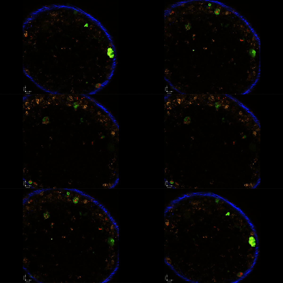
 The Toxoplasma parasite (in red) doesn’t need to infect an immune cell to alter its behavior, according to new Penn Vet research. Simply being injected with a package of proteins by the parasite (indicated by cells turning green) is enough to change the host cells’ activity.
Toxoplasma gondii
 is best known as the parasite that may lurk in a cat’s litter box. Nearly a third of the world’s population is believed to live with a chronic
Toxoplasma infection. It’s of greatest concern, however, to people with suppressed immune systems and to pregnant women, who can pass the infection to their fetuses.
Toxoplasma’s “success,” scientists believe, owes in part to its ability to evade the immune response of its host, whichever warm-blooded vertebrate it has infected. Now a new study suggests the parasite employs a sophisticated manipulation to suppress that immune response.
The work, led by researchers from Penn’s School of Veterinary Medicine and the University of Arizona and published in the Journal of Experimental Medicine, shows that T. gondii parasites inject their host’s macrophages, a type of immune cell, with a protein that changes the activity of the macrophage itself, creating what is known as an M2 macrophage. Those changes, the team showed, rein in the response of T cells that are normally responsible for killing parasites.
 Dr. Christopher Hunter
“This is the first time that it’s been shown that injection alone is sufficient to drive the creation of M2 macrophages,” says Christopher A. Hunter, an immunologist at Penn Vet and senior author of the paper. “Pharmaceutical companies have been targeting the pathways that create M2 macrophages for a long time because they’re very important in wound healing, fibrosis, lung repair, and so on. But here’s a parasite that manages to target it perhaps more efficiently than pharma has been able to.”
As the ability of the parasite to affect the infected and injected host cell populations has an impact on the course of infection, Hunter hopes further studies will help define the underlying mechanism.
For T. gondii, infection is a careful balancing act. It wants to spread far and wide within a host—and eventually be passed on to other hosts—but protective immunity is needed to prevent the death of the host. Scientists have long known that macrophages are critical players in maintaining this balance.
Macrophages are the cells normally responsible for cleaning up infections by consuming foreign invaders. Around a decade ago, scientists found that they come in different “flavors.”
“Some macrophages are profoundly pro-inflammatory and kill pathogens; these are known as M1 macrophages.” Hunter says. “M2 macrophages are profoundly anti-inflammatory but are less able to kill parasites. So M1 macrophages induce inflammation, and M2s help clear it up.”
 Longfei Chen and David Christian, two members of Hunter’s lab, were co-lead authors on the publication.
Earlier work by other labs had shown that T. gondii can act on a family of proteins that in turn has an influence over whether a macrophage becomes an M1 or M2. The parasite turns off the host STAT1 protein, which drives M1 production, and turns on host STAT3 and STAT6, which had both been shown to support the creation of M2 macrophages. But the consequences of this M2 production have been unclear, so Hunter and colleagues, led by lab members Longfei Chen and David A. Christian, sought to deepen their understanding of the parasite’s influence.
Previous work had suggested that the parasite activates STAT3 and STAT6 through an enzyme, ROP16 and that the parasite injects in host cells in a “package” of 2-300 proteins, of which the function of most is unknown. A collaborator and coauthor, Anita A. Koshy of the University of Arizona, enabled the research team to focus on the consequences of ROP16 injection by creating a genetically engineered strain of T. gondii. When this strain of parasite injects proteins into a cell, that cell glows green.
“We had previously recognized that some cells only receive this injection; they’re not actively infected with parasites,” Koshy says. “So, we thought if we can look only at the cells that received the injection we could ask what the consequences of that injection are.”
Unexpectedly, the researchers found that this population of immune cells that were injected but not infected were quite prominent. Both in cell culture and in mice infected with the engineered strain of T. gondii, the scientists were able to isolate this population and examine which genes were turned on in the injected only cells.
“In an infected cell, you see nearly 2,000 genes are changed,” says Hunter. “But if they’re only injected, you still see about 600 genes changing in expression.”
These changes alone were enough to sway macrophages over to the M2 type and to suppress the activity of T cells that normally act to kill parasites.
Finally, to investigate the effect of ROP16 on the parasite itself and its ability to infect mice, Josh Kochanowsky of the University of Arizona engineered a ROP16-deficient strain of T. gondii.
“If you put these parasites in immune-deficient mice, they grow normally” says Christian, “but if you put them in immune competent mice you get a reduced amount of M2s and a reduced parasite burden. So, we’re seeing that taking away ROP16 leads to a more effective immune response.”
Next for the team is to further study this pathway to get more conclusive information about how the M2 macrophages that are caused by the injection of ROP16—or other proteins—are able to suppress the host T cell response.
And because M2 macrophages are key players in anti-inflammatory processes that cancers also exploit to avoid detection by the immune system, Hunter says it’s possible that studying these parasites could reveal new information about the general biology of these M2 macrophages.
“Maybe this will tell us a bit more about how macrophages associated with tumors and infection can suppress a T cell response,” he says.
Christopher A. Hunter is the Mindy Halikman Heyer Distinguished Professor of Pathobiology in the University of Pennsylvania School of Veterinary Medicine.
Longfei Chen was a graduate student in the Hunter lab at the University of Pennsylvania School of Veterinary Medicine.
David A. Christian is a senior research associate in the Hunter lab at the University of Pennsylvania School of Veterinary Medicine.
Anita A. Koshy is an associate professor in the University of Arizona College of Medicine.
Joshua A. Kochanowsky is a graduate student in the Koshy Lab at the University of Arizona College of Medicine.
Hunter, Chen, Christian, Koshy, and Kochanowky’s coauthors were Anthony T. Phan, Joseph T. Clark, Shuai Wang, Corbett Berry, and Daniel P. Beiting of Penn Vet; Jung Oh and David S. Roos of Penn’s Biology Department in the School of Arts and Sciences; and Xiaoguang Chen of Southern Medical University in China.
The study was supported by the National Institutes of Health (grants AI126042-01, AI125563, NS095994, and AI41158), Commonwealth of Pennsylvania, University of Arizona, BIO5 Institute, Cancer Research Institute, Robertson Foundation, and National Natural Science Foundation of China.