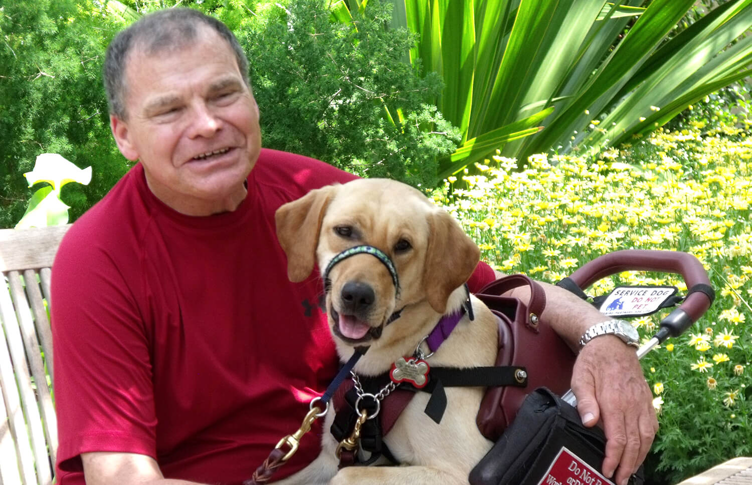
 Sheeba, at John Vagner's side
Sheeba never strays far from her owner John Vagner’s side. She’s his best bud and, as a diabetic alert dog trained by nonprofit Canine Partners for Life, his most astute signaler of a blood sugar drop or spike. The Labrador’s keen senses, particularly sight and smell, help her be the hard-working, loyal partner she is.
Every May, Vagner takes Sheeba for an eye check-up during the National Service Dog Eye Exam, an annual event sponsored by the American College of Veterinary Ophthalmologists (ACVO). Annually, participating veterinary ophthalmologists across the country provide free ocular exams to qualifying service dogs. Ryan Hospital is a regular participant in the ACVO program, offering eye exams to screen for conditions that could jeopardize a working dog’s vision.
When May 2015 rolled around, Vagner expected a routine screening for the then three-year-old dog. It was anything but. “We had no idea they’d find something,” he remembered. “Sheeba was fine—she played ball and just loved her life.”
Seeing What the Eye Can’t
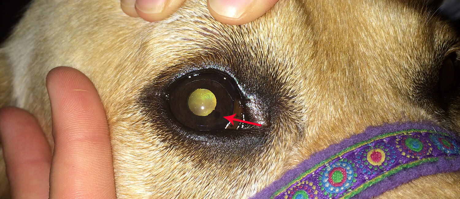
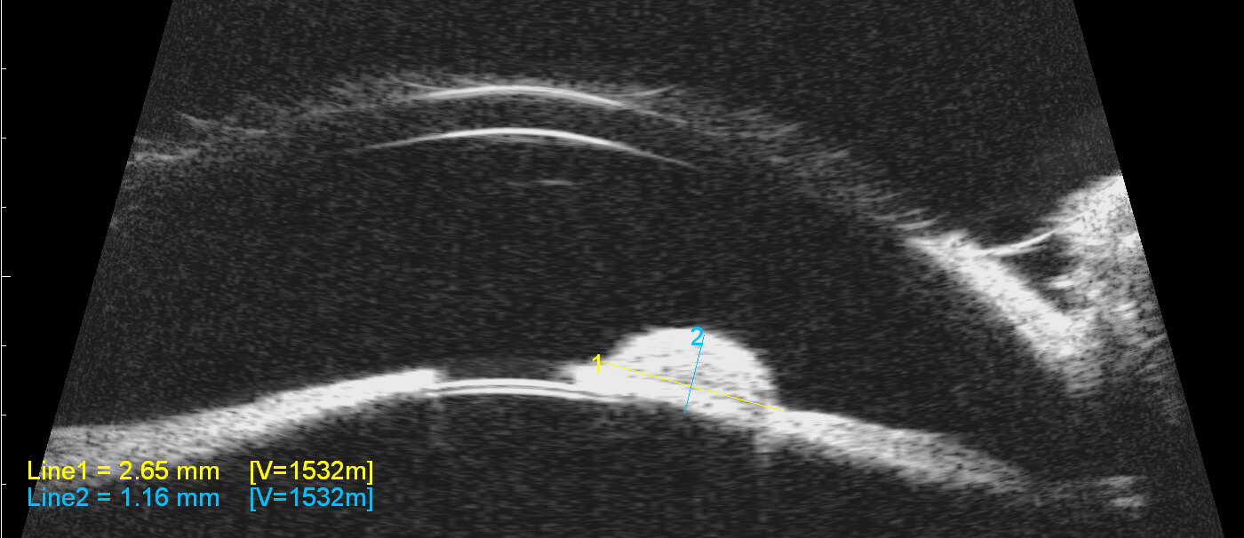
“The examining veterinary ophthalmologist near their home town of Oreland, Pennsylvania saw a dot on Sheeba’s iris and referred her to us for a closer look,” said Dr. Keiko Miyadera, Assistant Professor of Ophthalmology at Ryan Hospital. “We have a technique called ultrasound biomicroscopy—or UBM. It’s a noninvasive high-resolution imaging of the eye that enables us to pick up details we wouldn’t see in regular screening exams.” UBM involves numbing the eye and putting the probe directly on the cornea’s surface. This didn’t appear to phase Sheeba who “sat as still as a statue” for the exam.
Using the technology, Miyadera measured a 1.7 millimeter lesion on the iris in Sheeba’s right eye. It wasn’t large enough to bother the dog—at least not yet.
 Sheeba's lesion, the black dot at the red arrow, can be seen by the naked eye.
“It’s important to catch these masses way before the owners notice them with the naked eye or the dog starts to feel them,” Miyadera said. “By the time the animal starts to feel the effects of a growth, the lesion would have grown so much so it would have started to impact or injure the eye. Regular and early screenings are very important.”
Observe First, Remove Second
Lasering Sheeba’s mass was an option, but Vagner decided against the procedure at the time. Said Vagner, “We considered all the possibilities and decided to hold off—we didn’t want to put Sheeba through so much and instead chose to wait until the mass showed any sign of progression.”
The dog’s doctors supported the decision. “We typically tend not to jump right in and biopsy these masses in favor of less invasive approaches,” explained Dr. Brady Beale, a Clinical Instructor in Ophthalmology at Ryan Hospital. “Rather, we first go by clinical signs to gauge whether the spot is something benign and slow growing, such as a melanocytoma, or more aggressive, like a malignant melanoma. And even in there is a high suspicion of iris melanoma, the rate of metastasis is so low that we often elect monitoring as opposed to doing something invasive early on.”
Since 2015, Miyadera has examined Sheeba’s eye once a year. And every time the lesion’s margins have grown insignificantly. Until 2018.
“I measured a bit more growth during last year’s exam,” Miyadera explained. “So, we went ahead with laser therapy to reduce the size of the mass and decrease the change of further growth.”
 An ultrasound biomicroscopy image of Sheeba's eye before laser surgery showing a side view of the lesion
In December, a team supervised by Beale performed the procedure. “Immediately, we were able to reduce the mass by 50 percent, and it continued to shrink over the next few weeks after Sheeba was discharged,” Beale said.
The laser experience involves going under general anesthesia and receiving multiple ‘shots’ of lasers focused sharply on Sheeba’s iris lesion emitted from a headset worn by the clinical team. “She tolerated it beautifully. She was a great patient,” Beale said. Although, “it did take some coaxing to get her away from John—she’s so attached and always working for him!”
An emotional Vagner added. “Everything went as well as we could hope for. Doctors Miyadera and Beale were conscientious and caring—Penn Vet is a wonderful place.”